Ovarian Cancer Autoantibodies Reveal Hidden Immune Activation Pathways
A specific subset of ovarian tumors triggers natural immune proteins against cellular guardians, pointing toward improved early screening and tailored immunotherapies.
A specific group of ovarian cancer patients develops natural immune proteins against a crucial cellular guardian, pointing toward a fresh path for early detection and targeted immunotherapy. Researchers analyzing clinical samples discovered that these protective immune proteins, known as autoantibodies, target a famous tumor-suppressing protein called p53, but only when specific genetic mutations and protein buildups occur inside the malignant cells.
The investigation, published in Cancer, focused on 174 patients diagnosed with primary epithelial ovarian, peritoneal, and fallopian tube cancers within a comprehensive Danish medical cohort. Out of the entire group, 135 patients had a particularly aggressive and prevalent form of the disease called high-grade serous carcinoma. Elevated levels of immunoglobulin G anti-p53 autoantibodies showed up exclusively in 30 patients, and every single one of them carried this specific high-grade serous carcinoma diagnosis.
Why Only Certain Tumors Trigger Anti-p53 Immune Responses
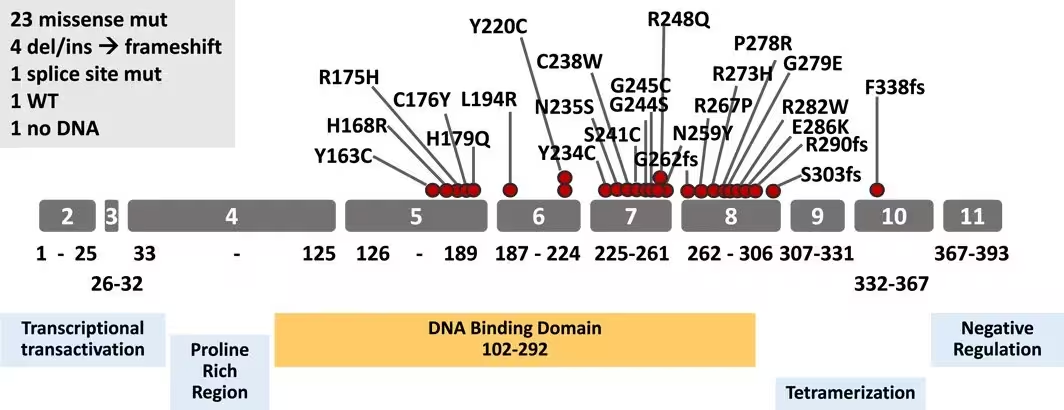
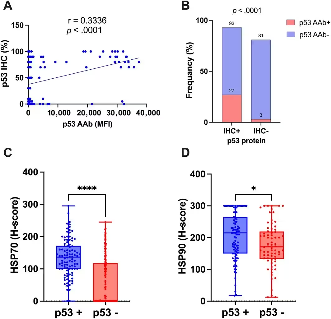
Cellular guardians normally work to prevent abnormal cell growth, but they frequently undergo damaging alterations in advanced malignancies. The team found that 97 percent of the patients who produced these p53 autoantibodies also harbored mutations in the TP53 gene, with 93 percent of those genetic flaws localized strictly within the DNA-binding domain of the protein. Furthermore, 89 percent of these antibody-positive tumor tissue samples displayed severe overexpression of the p53 protein under microscopic examination.
Molecular chaperones inside the cells appear to play a vital stabilizing role in this process. The analysis revealed that cytoplasmic levels of heat-shock proteins Hsp70 and Hsp90 were significantly elevated in tumors that tested positive for high p53 accumulation. Researchers suggest these chaperone molecules help stabilize the abnormal proteins and assist in presenting them to the immune system, thereby facilitating autoantibody production.
What Competition Assays Reveal About Target Epitopes
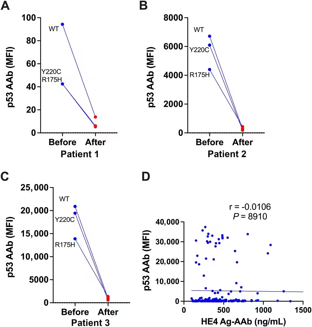
Medical science has long debated whether immune systems generate these autoantibodies in response to mutant variants or normal, wild-type protein structures. To answer this, investigators conducted competitive binding assays by pre-absorbing patient serum samples with normal wild-type p53 protein before testing them against specific mutant variants found in those patients. The absorption removed more than 85 to 98 percent of the antibody reactivity against the mutant proteins.
These absorption experiments indicate that the autoantibodies primarily recognize shared structural regions on normal, wild-type p53 rather than newly formed mutant sequences. Consequently, while gene mutations are necessary to trigger the immune reaction, the body responds to the sheer abundance of the protein rather than unique mutated shapes alone. The study also checked for cross-reactivity with human epididymis protein 4 antigen complexes and found no correlation, confirming that the immune production remains strictly specific to p53.
How an Active Tumor Microenvironment Shapes Immunity
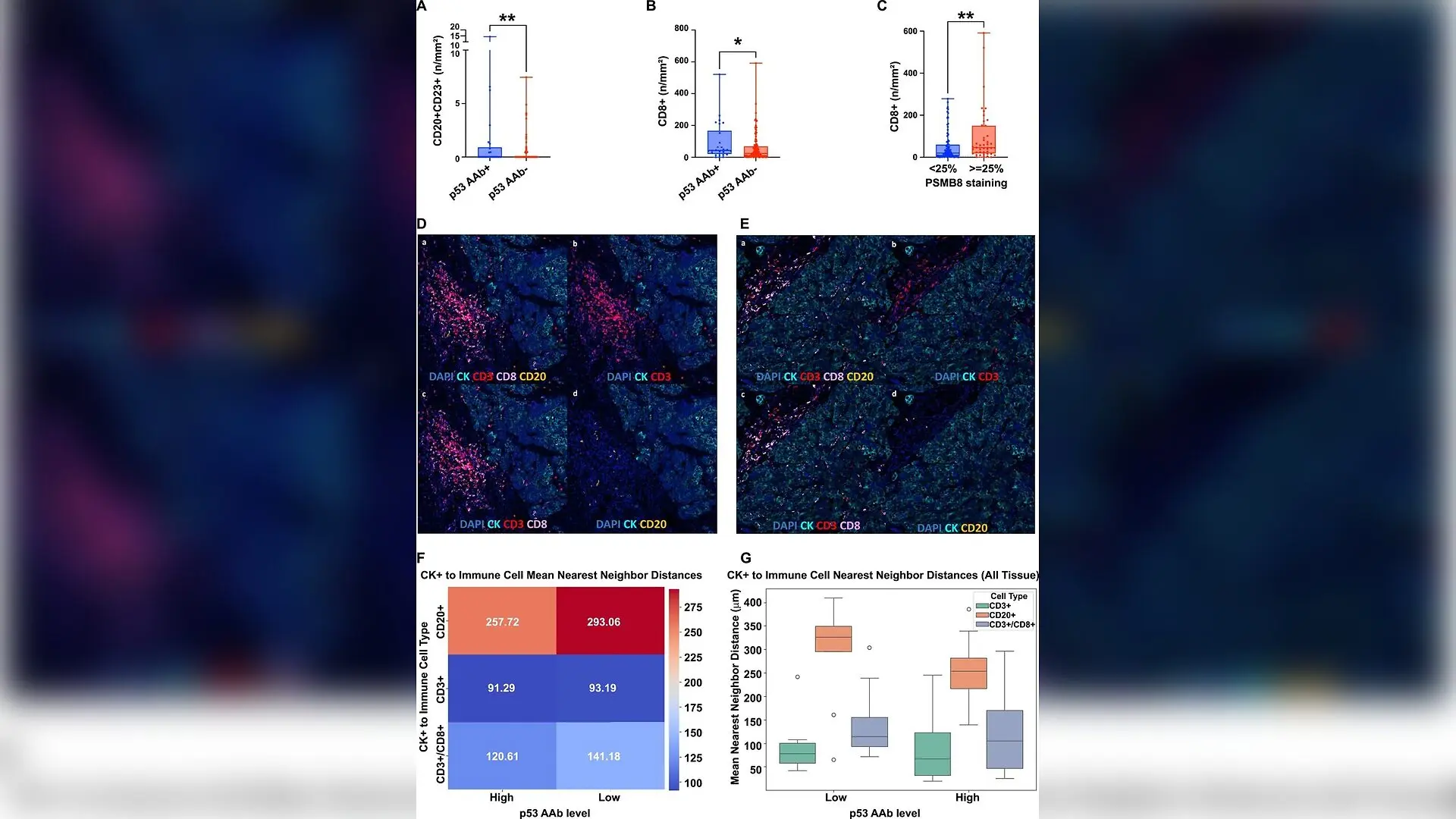
Patients who generated these p53 autoantibodies exhibited a distinctly active cellular neighborhood surrounding their tumors. Multiplex immunofluorescence staining showed that elevated autoantibody levels correlated strongly with heavy infiltration of CD20-positive and CD23-positive B cells, alongside cytotoxic CD8-positive T cells. Closer spatial measurements demonstrated that immune cells sat significantly closer to malignant cells in these specific patients.
Additionally, high expression of PSMB8, a key component of the immunoproteasome responsible for processing cellular antigens for immune presentation, tracked closely with increased cytotoxic T-cell counts. Despite this heightened cellular activity, the presence of these immune markers did not translate into a statistically significant improvement in overall or progression-free survival rates for the patients. This outcome implies that while the immune system recognizes the danger, the local microenvironment remains functionally constrained.
What Lies Ahead for Ovarian Cancer Biomarkers and Vaccines
Detecting ovarian malignancies early remains one of modern oncology’s greatest hurdles, as two-thirds of patients are diagnosed only after the disease reaches advanced stages. Previous screening data indicated that p53 autoantibodies can appear in patient blood samples months or even years before standard clinical markers like cancer antigen 125 register an elevated reading. Integrating these autoantibodies into multi-marker diagnostic panels could dramatically improve early detection timeframes.
On the therapeutic front, identifying patients who naturally mount a p53-directed immune response could help clinicians select candidates for specialized immunotherapies. Clinical trials combining p53-targeting vaccines with immune checkpoint inhibitors have already shown encouraging signs of extending progression-free survival in a fraction of advanced cases. Pinpointing individuals with pre-existing autoantibodies and active cellular infiltration may finally unlock higher response rates for these promising treatments.
The research was published in Cancer on November 15, 2026.
This article has been fact checked for accuracy, with information verified against reputable sources. Learn more about us and our editorial process.
Last reviewed on .
Article history
- Latest version
Reference(s)
- Qiu, Cuipeng., et al. “Biologic and immunologic characteristics of ovarian cancers that are associated with p53 autoantibodies.” Cancer, vol. 132, no. 22, November 13, 2026 Wiley, doi: 10.1002/cncr.70672. <https://doi.org/10.1002/cncr.70672>.
Cite this page:
- Posted by Priya Sharma