Microfluidic Chip Recreates Living Human Skin Dynamics in Real Time
A new microphysiological platform combines single-cell sequencing and live 3D imaging to capture how human skin cells divide, migrate, and stratify.
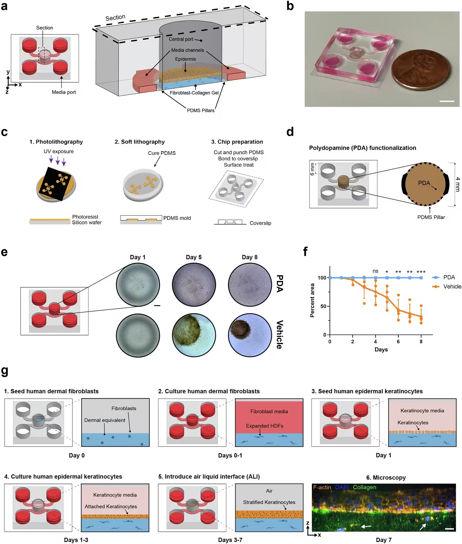
Engineered skin models often struggle to replicate the complex mechanics and living dynamics of human tissue, leaving researchers with limited ways to study how skin forms or where it fails in diseases like psoriasis. To overcome these hurdles, scientists at the University of California, San Francisco, designed a microfluidic platform called StrataChip, which grows living human epidermis over a media-perfused dermal tissue substitute within a microdevice.
The new device solves a persistent problem in lab-grown skin models, which is the tendency of collagen hydrogels to shrink and warp over time as fibroblasts pull on them. By coating the inner chambers with polydopamine, a sticky polymer that anchors the gel, the team prevented post-polymerization contraction entirely. This stability allows human epidermal keratinocytes to be seeded cleanly without delays or structural distortions, and the introduction of an air-liquid interface on the third day prompts rapid epidermal stratification within seven days.
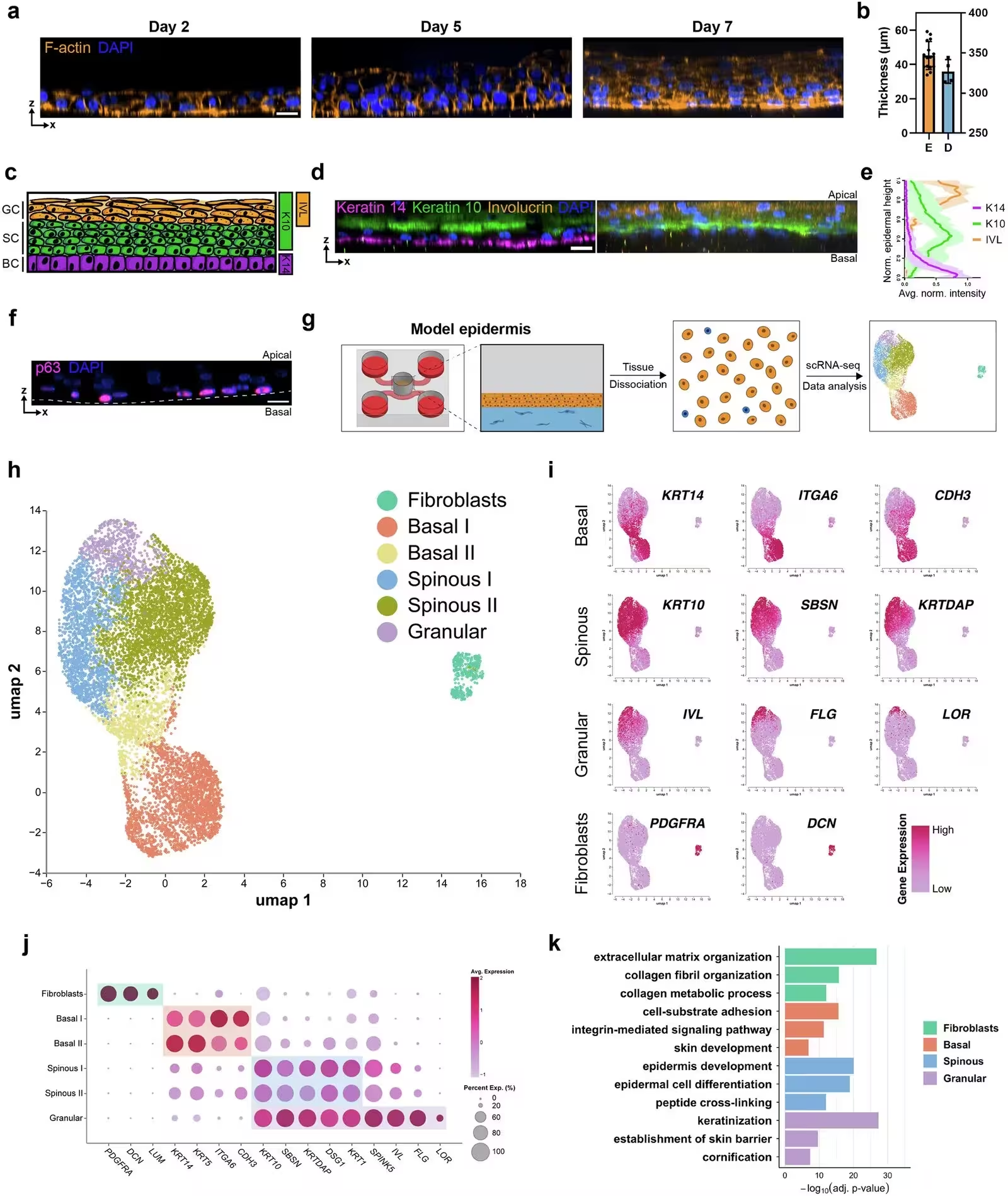
As detailed published in npj Biomedical Innovations, the resulting engineered tissue replicates the distinct architecture of real human skin, complete with basal, spinous, and granular layers. High-resolution confocal microscopy and single-cell RNA sequencing confirmed that the platform successfully develops characteristic cell markers, including keratin 14 in the basal layer and involucrin in the outer layers.
How Single-Cell Sequencing Revealed Hidden Skin Subpopulations
When the researchers isolated and sequenced thousands of individual cells from the lab-grown tissue, they uncovered unexpected cellular diversity within the layers. Automated clustering divided the basal keratinocytes into two distinct subpopulations, with one group specialized in cell-substrate adhesion and another showing elevated stress-associated keratins and early suprabasal markers. This transcriptional mapping highlights how individual cells transition from proliferative stem-like states toward terminal differentiation.
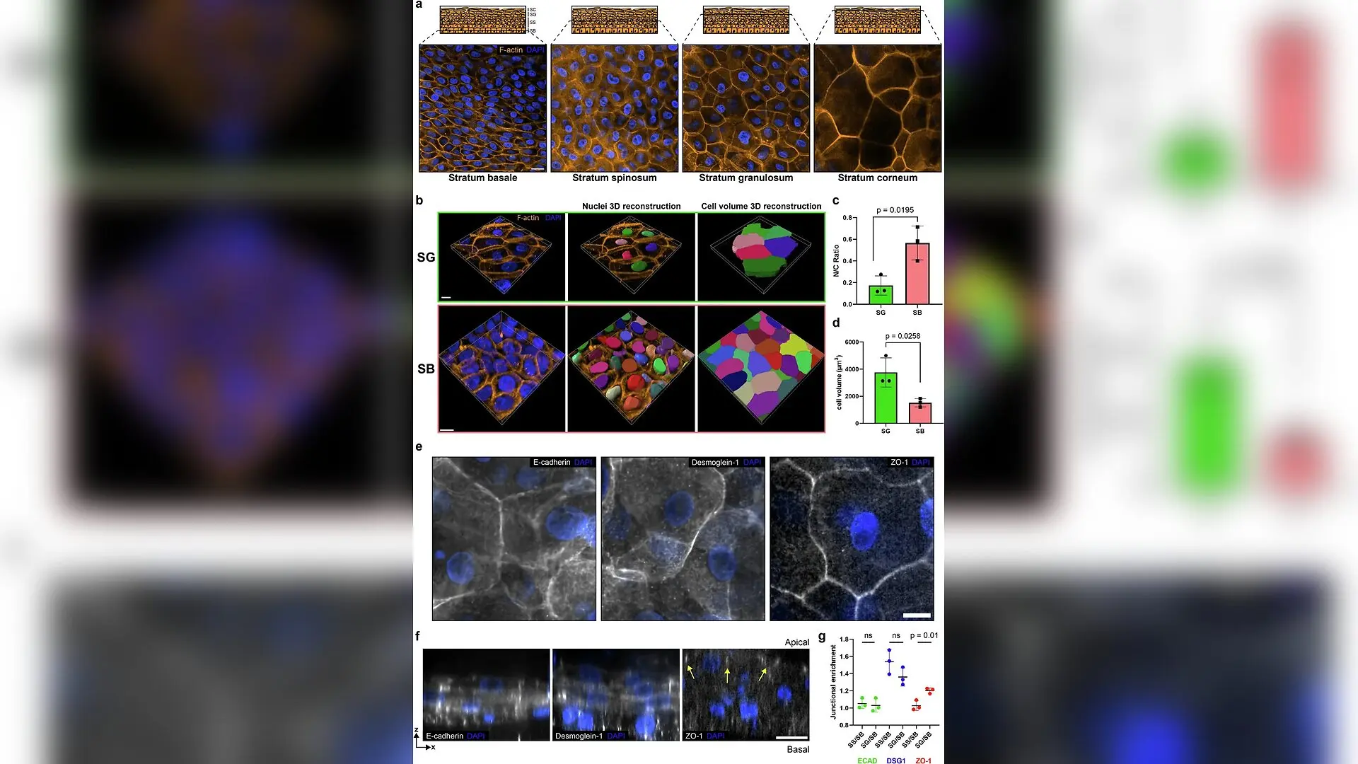
The platform also mirrored the spatial organization of crucial adhesion molecules found in natural skin. E-cadherin was distributed evenly across all layers to maintain tissue integrity, while desmoglein-1 concentrated heavily in the upper spinous and granular compartments, aligning with mature desmosome formation. Tight junction proteins such as ZO-1 restricted themselves strictly to the outermost living cells, confirming that the tissue builds a proper functional barrier.
Why Real-Time 3D Imaging Changes the Study of Morphogenesis
Traditional organotypic cultures are typically fixed at single time points, making it difficult to watch cellular events unfold in real time. By combining fluorescent live-cell probes with advanced spinning-disk confocal and oblique plane microscopy, the team captured active morphodynamic behaviors directly inside the living 3D tissue.
The live imaging sessions recorded basal keratinocytes detaching and migrating upward into suprabasal layers through a process called delamination, as well as asymmetric cell divisions where one daughter cell retained matrix contact while the other moved outward. Researchers also observed nuclear degradation and DNA leakage in apically located cells, providing a direct visual window into cornification and terminal differentiation.
What Limitations Remain and Where the Technology Goes Next
Despite its detailed structural and genetic fidelity, the study notes that the current platform does not yet fully characterize the functional properties of the physical epidermal barrier or underlying dermal mechanics, which will require further physiological validation. Furthermore, long-term cultures extending past fourteen days may introduce stability challenges that require additional stromal cell optimization.
Even with these boundaries, the ability to merge a microphysiological model with single-cell transcriptomics and real-time live imaging opens new paths for disease research. Scientists can now introduce specific genetic mutations or inflammatory cues into the system to watch how cellular mechanics and gene regulation fracture in conditions like basal cell carcinoma, autoimmune blistering disorders, and chronic dermatitis.
The research was published in npj biomedical innovations on November 12, 2026.
This article has been fact checked for accuracy, with information verified against reputable sources. Learn more about us and our editorial process.
Last reviewed on .
Article history
- Latest version
Reference(s)
- Amakor, Justin K.., et al. “StrataChip: a microphysiological system to capture dynamic keratinocyte fate and mechanical transitions during human epidermal morphogenesis.” npj Biomedical Innovations, vol. 3, no. 1, November 12, 2026 Springer Science and Business Media LLC, doi: 10.1038/s44385-026-00124-3. <https://doi.org/10.1038/s44385-026-00124-3>.
Cite this page:
- Posted by Rohan Kumar