AI Scans Soles to Screen for Leg Alignment and Knee Risk
A novel multimodal deep learning approach uses weight-bearing 3D foot scans to classify lower-limb alignment without radiation.
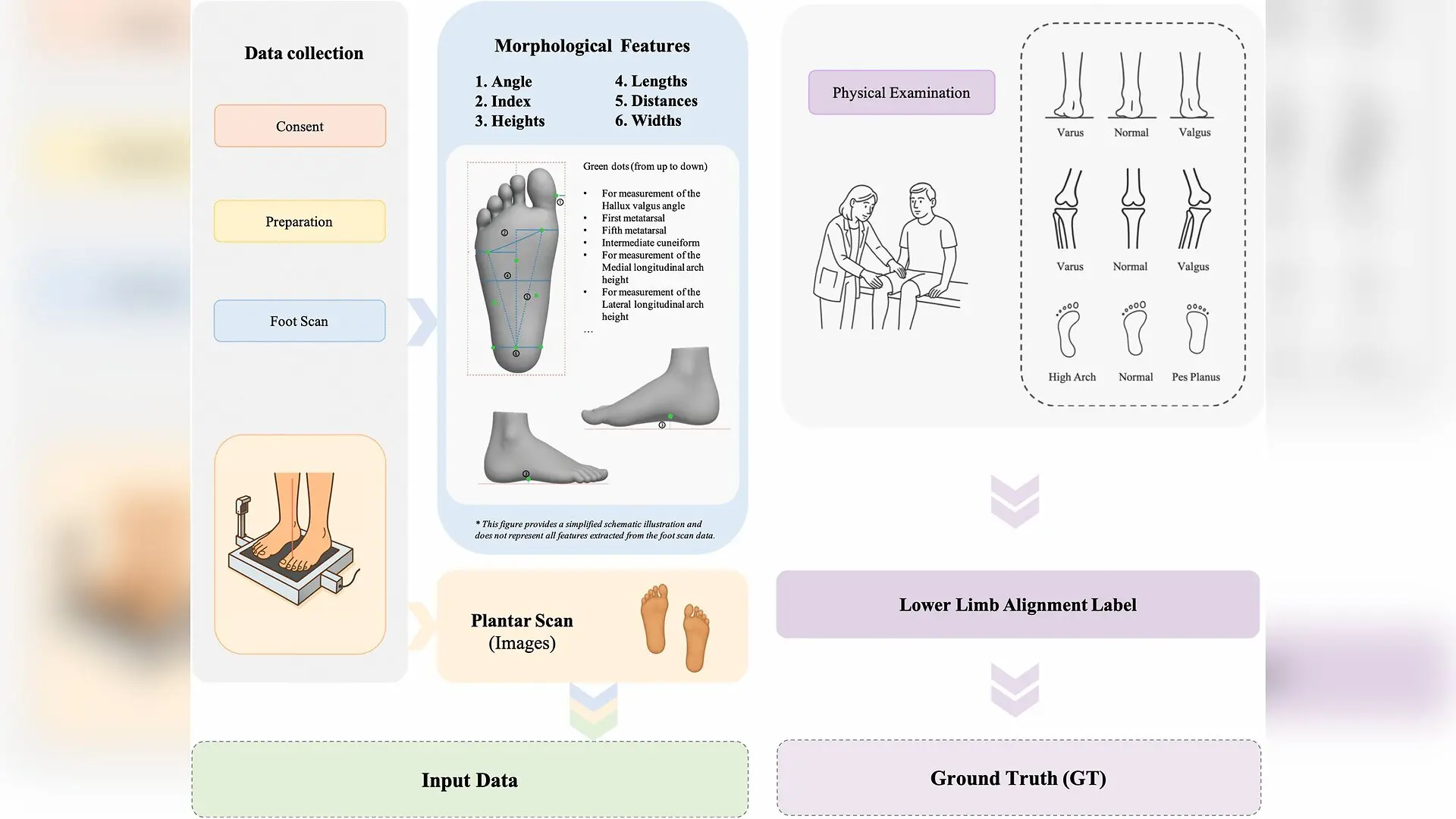
Routine screening for lower-limb malalignment typically requires exposure to ionizing radiation through radiography or a specialist clinical examination, which limits its availability for early detection of conditions like knee osteoarthritis and plantar fasciitis. Researchers have investigated whether a simple weight-bearing 3D foot scan can serve as a non-invasive computational screening tool for proximal alignment across the entire kinetic chain, running from the foot up to the hip. The work, published in PLOS Digital Health, outlines an attention-based multimodal deep learning framework designed to classify arch types, hindfoot alignment, and frontal-plane knee alignment from plantar images and geometric measurements.
How Artificial Intelligence Combines Foot Scans and Geometry
The team of researchers from institutions including Shanghai Jiaotong University School of Medicine established a cross-sectional dataset of 167 participants, spanning ages 6 to 60 years, who underwent static full weight-bearing 3D foot scanning. Each scan yielded a detailed RGB plantar image alongside 16 quantitative morphological features, such as medial arch height, forefoot width, hallux valgus angle, and heel width. Clinical experts independently evaluated and labeled the feet for arch type, hindfoot alignment relative to the tibial axis, and knee alignment.
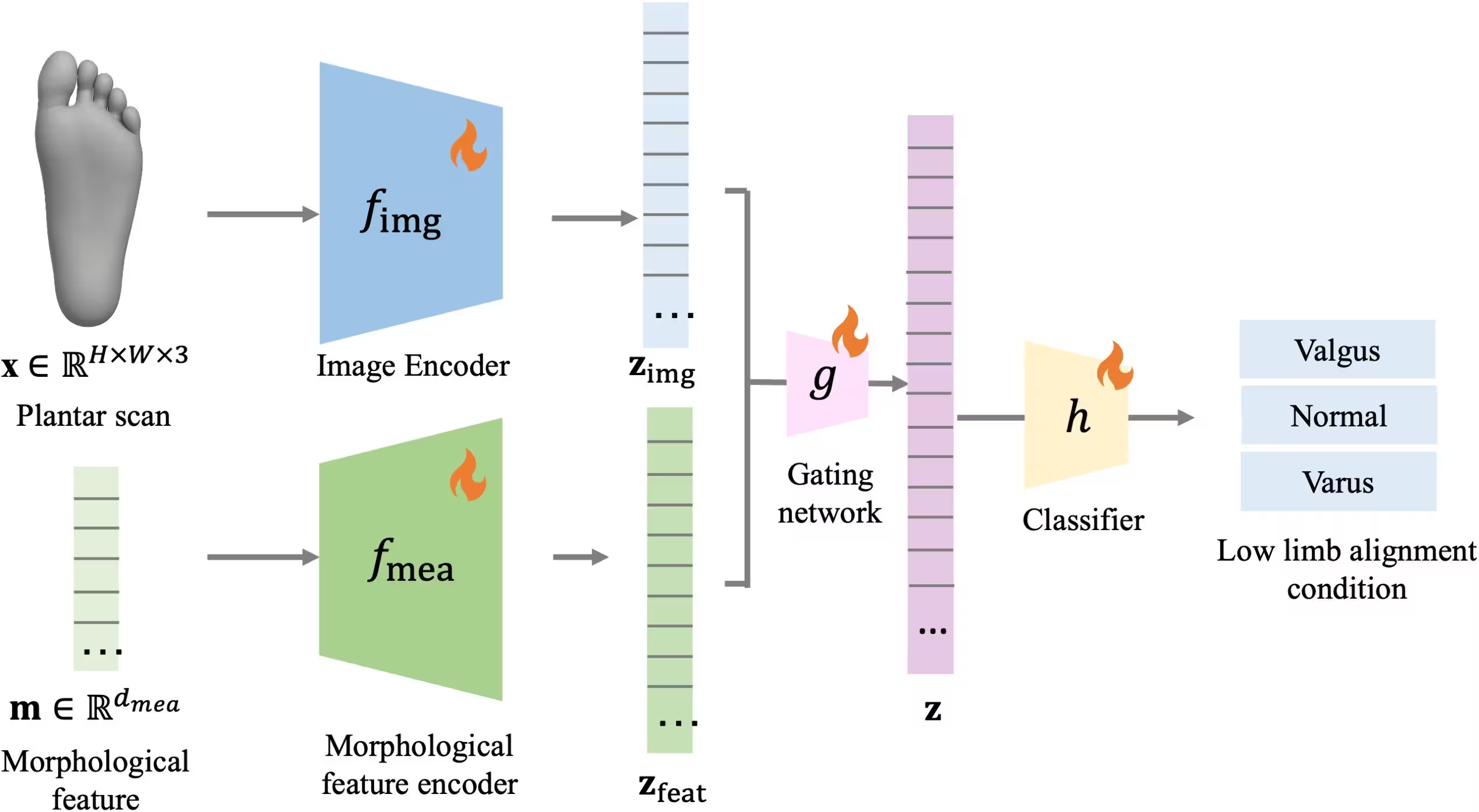
To process this data, the investigators developed a dual-encoder deep learning model featuring an image feature encoder paired with a morphological feature encoder. A feature-wise attention fusion module then adaptively combined the visual embeddings with the numerical measurements before passing them to a classification network. The architecture was benchmarked across four major visual backbone families, namely Inception, ResNet, EfficientNet, and the Vision Transformer, while being compared against morphology-only machine learning models and image-only configurations.
What the Multi-Class Classification Performance Shows
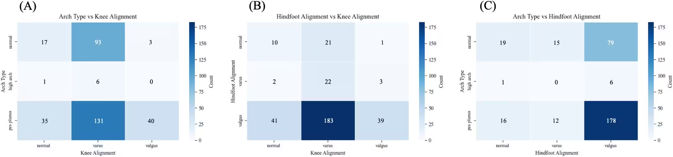
Evaluated on a held-out test set using bootstrap resampling, the proposed multimodal framework demonstrated strong performance, particularly for arch type classification. Models using the Inception V3 backbone with attention-based fusion achieved an overall accuracy of 89 percent with an area under the ROC curve of 0.98 for arch type. For hindfoot alignment, the fusion model reached an accuracy of 81 percent, though the authors noted a heavy class imbalance where valgus accounted for over 80 percent of the dataset. Knee alignment classification proved to be the most challenging task, achieving an accuracy of 76 percent with the Inception attention-fusion setup.
Class activation mapping techniques, including Grad-CAM, Score-CAM, and Layer-CAM, were applied to visualize which plantar regions drove the model decisions. For arch type predictions, the activation heatmaps consistently focused on the medial longitudinal arch area. In knee alignment tasks, the models highlighted the central plantar area and the region beneath the first metatarsophalangeal joint, aligning with known biomechanical loading shifts associated with frontal-plane knee malalignment.
Limitations and Next Steps for Digital Screening
Despite promising results, the study authors emphasize several important limitations to keep in mind. The dataset was relatively modest and drawn from a single clinical centre, and the broad age range introduced developmental heterogeneity that could not be fully stratified given the sample size. Furthermore, alignment labels relied on clinical physical examination rather than full-leg standing radiography, which remains the gold standard for coronal-plane measurements. The static nature of the scans also means dynamic muscular compensations and ground reaction forces during locomotion were not captured.
The researchers note that while the attention-based fusion framework offers an explainable and scalable method for preliminary alignment assessment, its clinical translation will require validation in larger, multi-centre cohorts with external testing. Future work will need to incorporate dynamic modalities, such as plantar pressure measurements or gait analysis, and assess longitudinal outcomes to better understand how foot morphology relates to progressive musculoskeletal conditions.
The research was published in PLOS Digital Health on November 11, 2026.
This article has been fact checked for accuracy, with information verified against reputable sources. Learn more about us and our editorial process.
Last reviewed on .
Article history
- Latest version
Reference(s)
- Ni, Simin., et al. “Development and internal validation of a multimodal deep learning model for screening-level lower-limb alignment using plantar 3D foot scans.” PLOS Digital Health, vol. 5, no. 11, November 11, 2026, pp. e0001802 Public Library of Science (PLoS), doi: 10.1371/journal.pdig.0001802. <https://doi.org/10.1371/journal.pdig.0001802>.
Cite this page:
- Posted by Robert Brown