Wrinkled MXene Coatings Guide Skeletal Muscle Cell Alignment
Engineered microscale topographies on scalable multilayer films promote myoblast fusion and accelerate the structural maturation of muscle tissue.
Engineered muscle tissue constructs require precise topographical cues to replicate the aligned architecture of native skeletal muscle, yet traditional microfabrication techniques remain costly and technically demanding. A team of researchers has developed a mold-free, scalable manufacturing strategy using wrinkled titanium carbide MXene multilayer coatings to successfully guide myoblast alignment, enhance cell proliferation, and accelerate the formation of mature multinucleated myotubes.
How Wrinkled Surface Geometries Guide Developing Muscle Cells
Mature skeletal muscle consists of densely packed, multinucleated myofibers aligned along a shared axis to enable effective contractile function. When substantial tissue loss occurs due to trauma or disease, the body struggles to repair the damage because mature myonuclei cannot divide and the population of resident stem-like satellite cells is extremely sparse, falling below 5 percent. Researchers have long sought to create engineered biomaterials that provide contact guidance to direct cells into organized arrays. Conventional approaches such as photolithography and nanoimprinting require cleanroom facilities, photomasks, and complex master molds, which severely limit clinical translation.
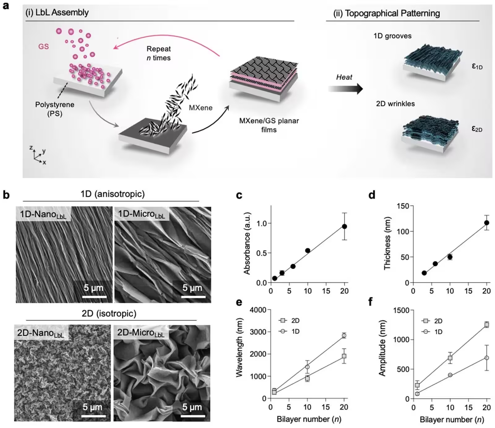
The research team addressed this limitation by designing a bottom-up fabrication method combining layer-by-layer assembly with uniaxial thermal shrinkage. First, investigators deposited alternating nanoscale layers of Ti3C2Tx MXene nanosheets and gentamicin sulfate onto pre-strained polystyrene substrates. Upon heating above the glass transition temperature of the polymer, the substrate shrank and forced the stiff MXene coating to buckle into periodic ripples. By controlling the number of deposited bilayers from 1 to 20, the team tuned the resulting surface topographies from nanoscale dimensions to microscale wavelengths measuring up to 2,830 nanometers. The study is published in Biomaterials Science as an open-access report detailing how these tunable surfaces interact with murine C2C12 myoblasts.
Evaluating Alignment and Proliferation on Nanoscale Versus Microscale Ridges
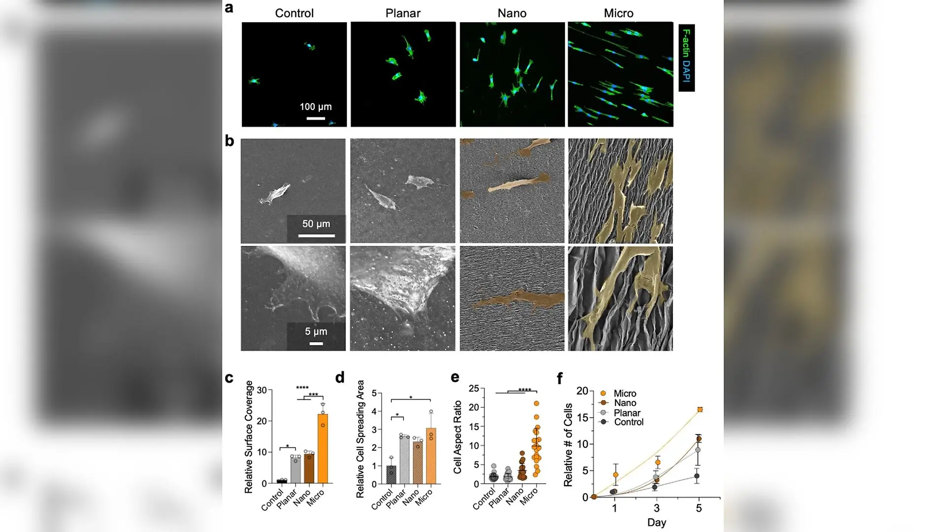
To test how different physical dimensions influence cell behavior, the authors compared flat control surfaces, unwrinkled planar MXene coatings, nano-wrinkled surfaces, and micro-wrinkled surfaces. Fluorescence and scanning electron microscopy revealed that cells cultured on micro-wrinkled coatings developed highly ordered, spindle-like morphologies oriented precisely parallel to the wrinkle axes. In contrast, cells on planar and nano-wrinkled substrates spread randomly in polygonal shapes with disorganized actin filament networks. Quantitative image analysis demonstrated that cell coverage expanded more than 22-fold on micro-wrinkled surfaces compared to untreated controls, while cell spreading area and aspect ratio also reached their maximum values on the micro-structured topographies.
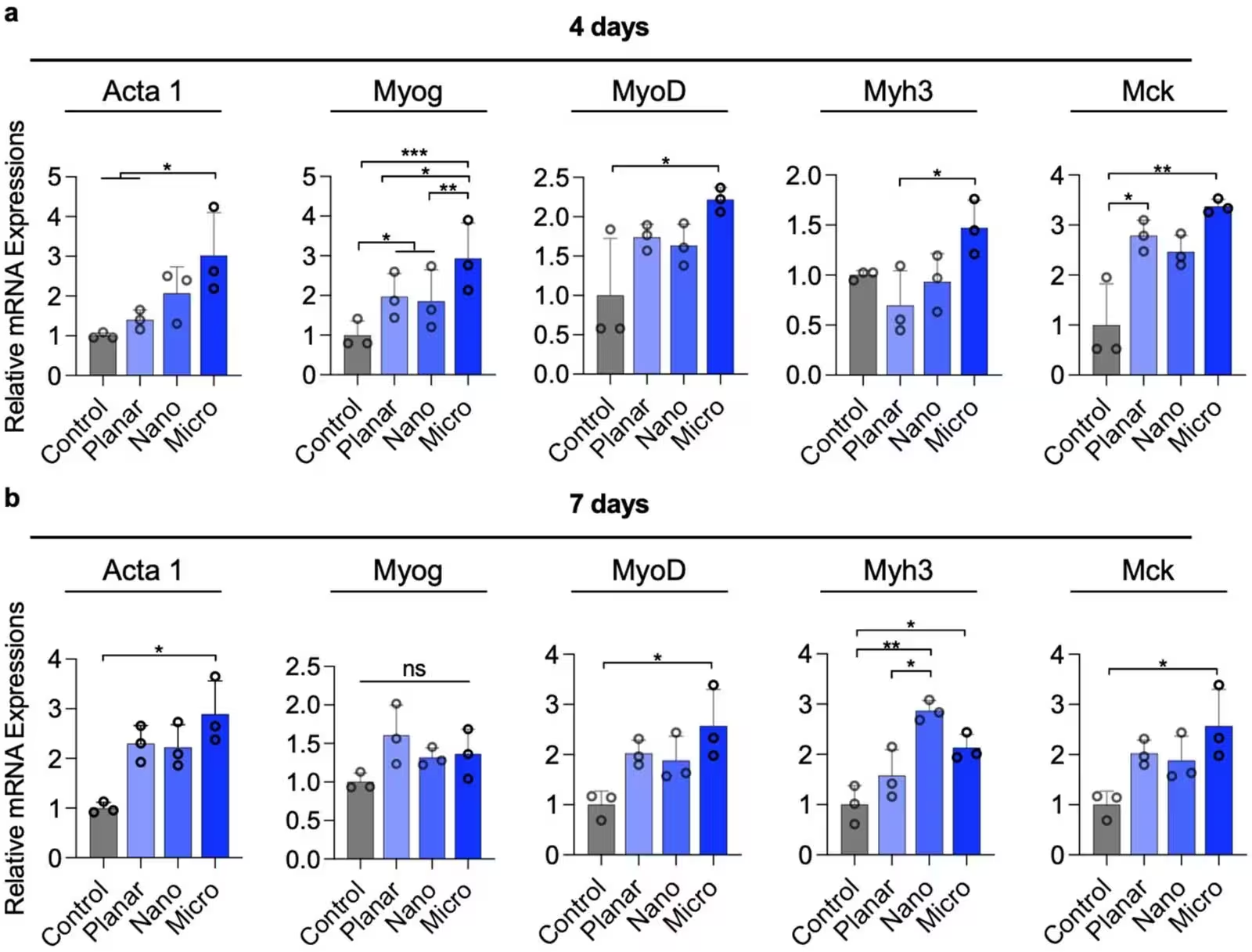
Proliferation assays confirmed that micro-wrinkled surfaces supported robust cell expansion over a five-day period. Furthermore, gene expression profiling via quantitative PCR showed that after four days in differentiation medium, myoblasts on micro-wrinkled substrates exhibited significant upregulation of key myogenic regulators such as MyoD and Myogenin, alongside structural markers including alpha-skeletal actin and muscle creatine kinase. This coordinated transcriptional activation indicated that microscale topographical cues promote both early lineage commitment and subsequent differentiation far more effectively than flat or nanoscale alternatives.
Advancing Structural Maturation and Myotube Fusion
Effective muscle regeneration requires individual precursor cells to fuse into elongated, multinucleated myotubes that organize functional contractile machinery known as sarcomeres. Cultures grown on micro-wrinkled MXene films formed abundant, highly aligned myotubes extending over 385 micrometers in average length. The myotube fusion index reached over 52 percent on these micro-structured substrates, significantly outperforming flat control surfaces and unwrinkled coatings. Immunostaining for sarcomeric alpha-actinin revealed periodic striations along the myotubes, indicating advanced myofibrillar maturation and well-organized sarcomeric units that were absent or poorly formed in less anisotropic groups.
The authors note several important limitations regarding their current platform. Although the wrinkled morphology remained stable for at least 14 days under simulated cell-culture conditions, the long-term chemical oxidation behavior of Ti3C2Tx MXene in biological media requires continued investigation. Additionally, all experiments were conducted on rigid polystyrene substrates that are substantially stiffer than native skeletal muscle tissue, meaning future studies will need to transfer these anisotropic MXene coatings onto compliant, elastic hydrogels or soft polymeric scaffolds that better match the mechanical microenvironment of living muscle.
Next Steps for Bioinspired Muscle Regeneration
By establishing a clear link between surface geometry and transcriptional regulation, this work provides an accessible and scalable manufacturing avenue for regenerative medicine. The ability to generate complex topographical cues without cleanroom infrastructure opens new possibilities for engineering functional biointerfaces. Subsequent developments may integrate electrical conductivity and bioactive modifications into the MXene platform to further improve tissue contractility, electrophysiological responsiveness, and long-term therapeutic efficacy in preclinical models of muscle injury.
The research was published in Biomaterials science on November 10, 2026.
This article has been fact checked for accuracy, with information verified against reputable sources. Learn more about us and our editorial process.
Last reviewed on .
Article history
- Latest version
Reference(s)
- Asadi Tokmedash, Mohammad., et al. “Tunable anisotropic nano/micro-topography aligns myotubes and accelerates maturation.” Biomaterials Science, January 1, 2026 Royal Society of Chemistry (RSC), doi: 10.1039/d6bm01020b. <https://doi.org/10.1039/d6bm01020b>.
Cite this page:
- Posted by Robert Brown