Bacterial Membrane Models Reveal Spontaneous Nanoscale Reorganization
Physical contact with supporting surfaces drives unexpected lipid segregation and domain formation in model prokaryotic membranes.
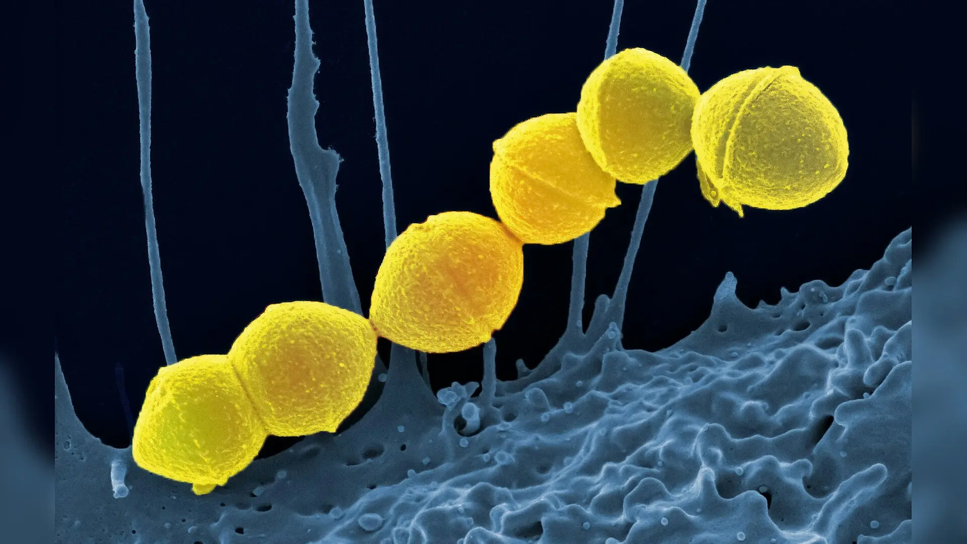
Bacterial cell membranes can spontaneously reorganize into distinct nanoscale patches within hours when placed on solid surfaces, driven purely by physical contact rather than active biological machinery. Researchers investigated this phenomenon by placing simplified lipid models of the Escherichia coli inner membrane onto various substrates, revealing a hidden structural plasticity that could fundamentally change how scientists view microbial cell walls.
Mimicking Bacterial Membranes Without Sterols
Unlike eukaryotic cells, which rely on cholesterol and complex sterols to regulate membrane fluidity and form functional microdomains known as lipid rafts, most prokaryotic membranes lack these molecules. Instead, bacteria survive through lipid diversity and homeoviscous adaptation, adjusting their lipid makeup to match environmental shifts. To isolate pure physical contact forces from complex biological signaling, researchers built a simplified binary model membrane containing 75 percent 1-palmitoyl-2-oleoyl-sn-glycero-3-phosphoethanolamine and 25 percent 1-palmitoyl-2-oleoyl-sn-glycero-3-phospho-(1′-rac-glycerol). This mixture mimics the natural phase transition temperature and mechanical properties of the E. coli inner membrane without relying on cardiolipin, a lipid known to naturally promote domain clustering.
The team tracked the physical behavior of these model membranes using high-resolution atomic force microscopy in liquid environments alongside differential scanning calorimetry. As published in Soft Matter, the experiments maintained the sample temperatures just a few degrees above their standard solution phase transition point. This setup allowed the team to capture the subtle ways in which supporting surfaces alter lipid organization over extended periods exceeding 24 hours.
Substrate Contact Triggers Spinodal Decomposition
When supported on an atomically flat mica surface, the lipid bilayers initially formed standard solid-ordered domains that grew slowly over the first hour. However, between one and three hours, the growth slowed down and reversed as thinner, fluid-disordered domains began appearing right inside the solid patches. This unexpected transformation resembled a spinodal decomposition, a spontaneous phase separation process that occurs without energy barriers.
The authors propose that water- and ion-mediated interactions between the membrane and the underlying solid substrate bias the passive flip-flop of lipids between the upper and lower leaflets. Because lipid flip-flop across a bilayer is normally energetically costly and slow without enzymatic help, the constraining presence of a surface appears to catalyze this redistribution. Over time, this asymmetry alters the local lipid composition, creating stable nanodomains with distinct thicknesses.
Shifting Timescales on Realistic Polymer Surfaces
To test whether surface texture alters this behavior, the team replaced the rigid mica with oxidized polydimethylsiloxane, a softer polymer cushion mimicking the mechanical properties of the peptidoglycan cell wall found naturally in bacteria. On this uneven surface, the entire reorganization process accelerated dramatically. The transition phases that took hours on mica occurred in less than thirty minutes on the polymer, demonstrating that surface topography and flexibility directly control the speed of membrane restructuring.
Furthermore, nanomechanical mapping using atomic force microscopy tips revealed that these newly formed nanodomains exhibited significantly different physical properties. The reorganized solid regions became noticeably stiffer and harder to puncture compared to the surrounding fluid areas, highlighting how physical crowding and support can drastically modify local membrane mechanics.
Broader Implications for Cellular Architecture
These findings suggest that physical contact with cellular structures like the cytoskeleton or the cell wall may actively shape bacterial membrane organization in living cells. While the experiments were conducted on in vitro model systems, the discovery points to a universal physical mechanism where surface proximity drives spontaneous lipid segregation. Understanding this contact-induced reorganization provides fresh context for how bacteria maintain mechanical integrity and handle osmotic pressure in changing environments.
The research was published in Soft matter on November 18, 2026.
This article has been fact checked for accuracy, with information verified against reputable sources. Learn more about us and our editorial process.
Last reviewed on .
Article history
- Latest version
Reference(s)
- Tormena, Nicolo., et al. “Contact-induced molecular reorganisation in a model Escherichia coli lipid membrane.” Soft Matter, vol. 22, no. 44, January 1, 2026, pp. 7395-7407. Royal Society of Chemistry (RSC), doi: 10.1039/d6sm00923a. <https://doi.org/10.1039/d6sm00923a>.
Cite this page:
- Posted by Hassan Raza