AI Scan Analysis Challenges Assumptions About Muscle Size and Back Pain
Evaluating over 10,000 computed tomography scans reveals that bulk trunk muscle size does not differ in back pain patients, pointing researchers toward fat-to-muscle ratios.
Deep learning analysis of more than 10,000 routine abdominal computed tomography scans reveals that overall trunk muscle size is not significantly smaller in individuals with low back pain, challenging long-held assumptions about core muscle wasting. The study, published in PLOS Digital Health, utilized automated image segmentation tools to evaluate body composition across 23,508 unique patients treated at a tertiary care center between 2013 and 2018.
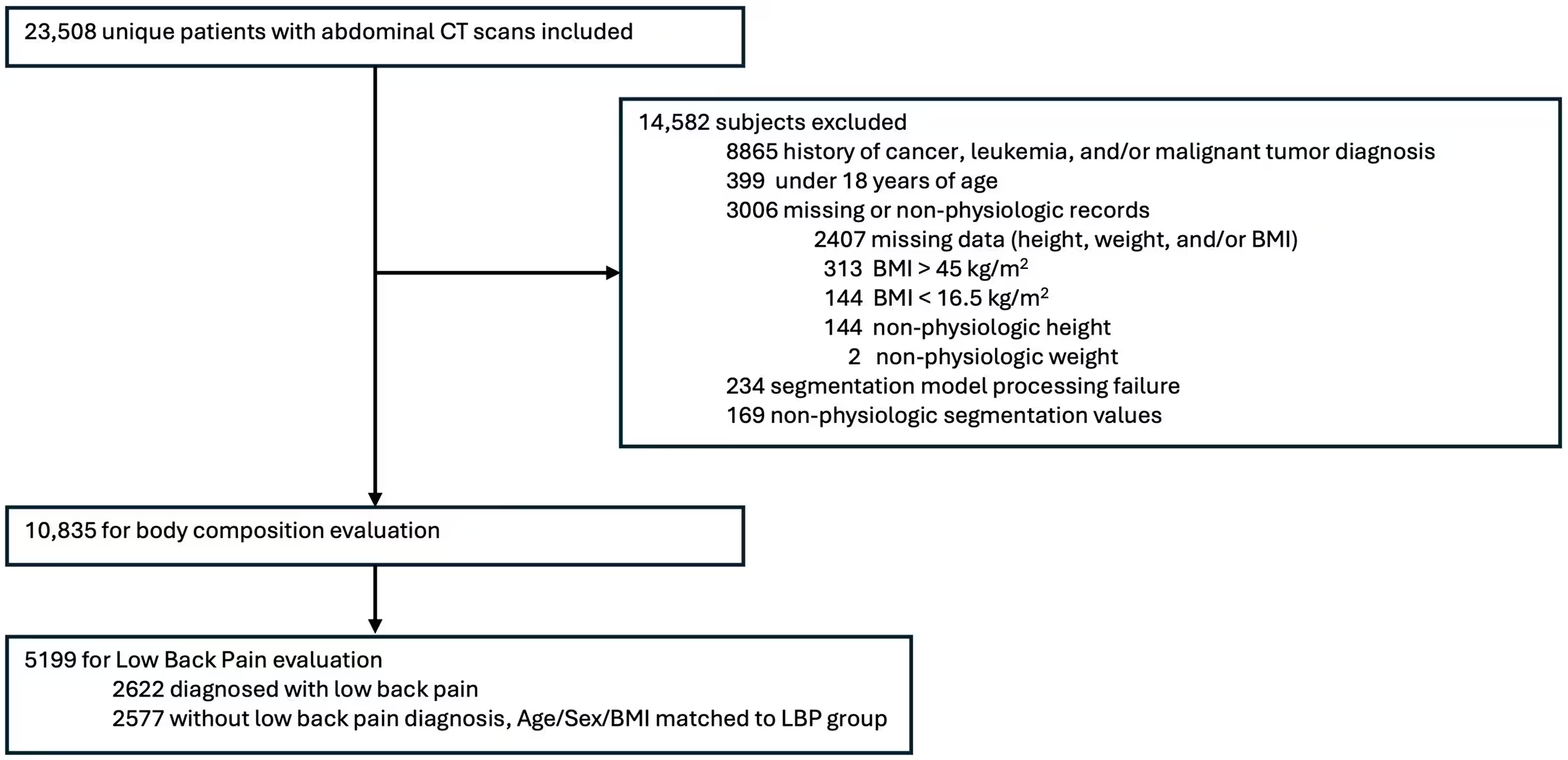
While previous clinical investigations into musculoskeletal health and back discomfort often relied on small cohorts of roughly 100 participants, this retrospective study scaled up the approach significantly. The researchers applied a validated open-source deep learning model known as Comp2Comp to examine cross-sectional areas of skeletal muscle, visceral adipose tissue, and subcutaneous adipose tissue at the level of the third lumbar vertebra. Out of the initial patient pool, 10,835 subjects met the strict inclusion criteria for the broader body composition normative analysis, while 2,622 patients diagnosed with low back pain were matched against 2,577 control subjects by age, sex, and body mass index.
How Artificial Intelligence Unlocked Decades of Medical Imaging Data
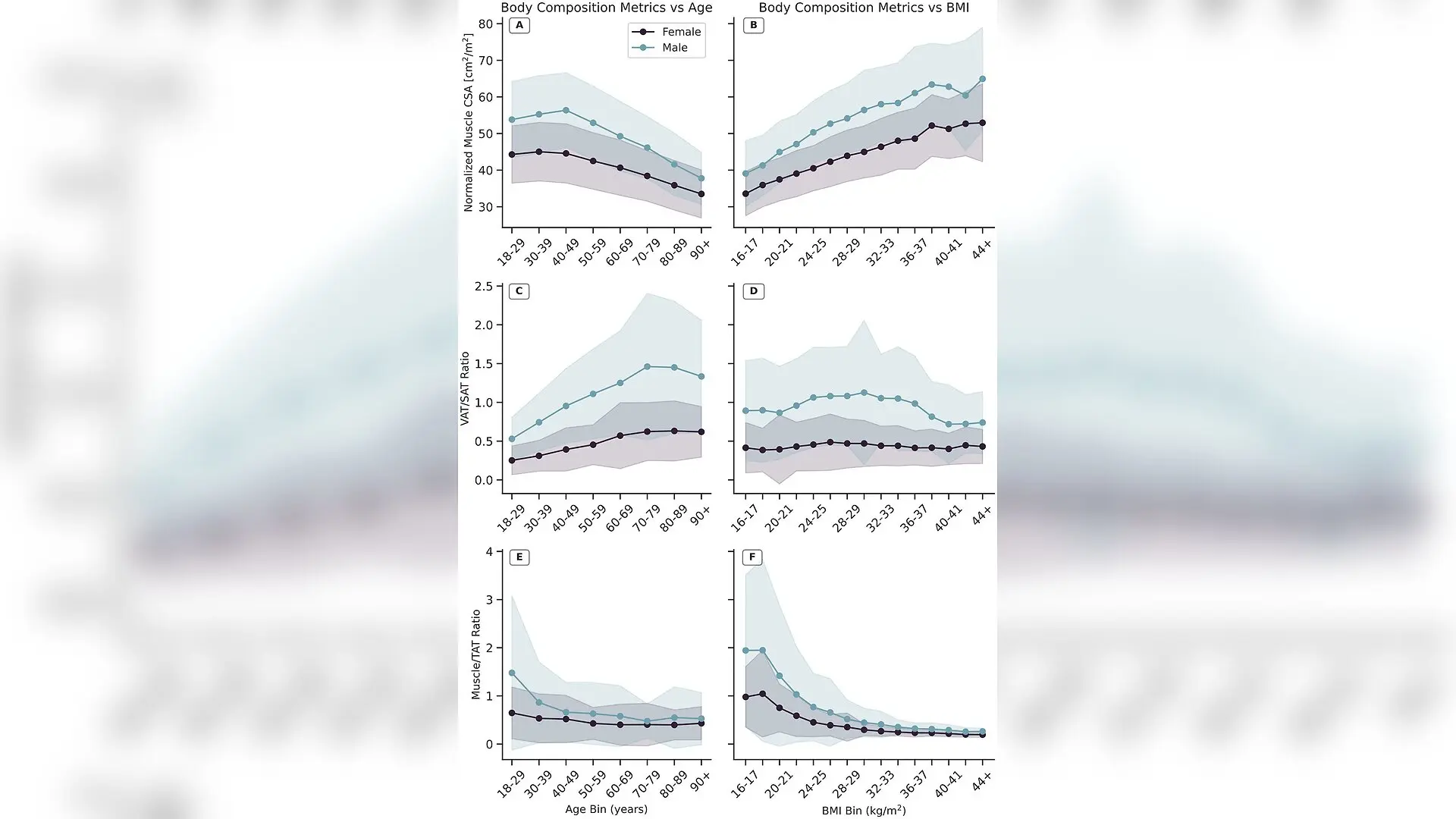
Routine medical imaging generates millions of diagnostic scans annually, yet these digital records typically remain archived and unused after initial clinical care. By deploying automated neural networks capable of segmenting tissue types with errors under two percent, the research team bypassed the exorbitant time and monetary costs traditionally associated with prospective epidemiological studies. To validate this opportunistic methodology, the authors first tested whether their dataset could reproduce established biological trends, such as the natural decline of height-squared normalized muscle cross-sectional area with advancing age and the steady increase of the visceral-to-subcutaneous fat ratio.
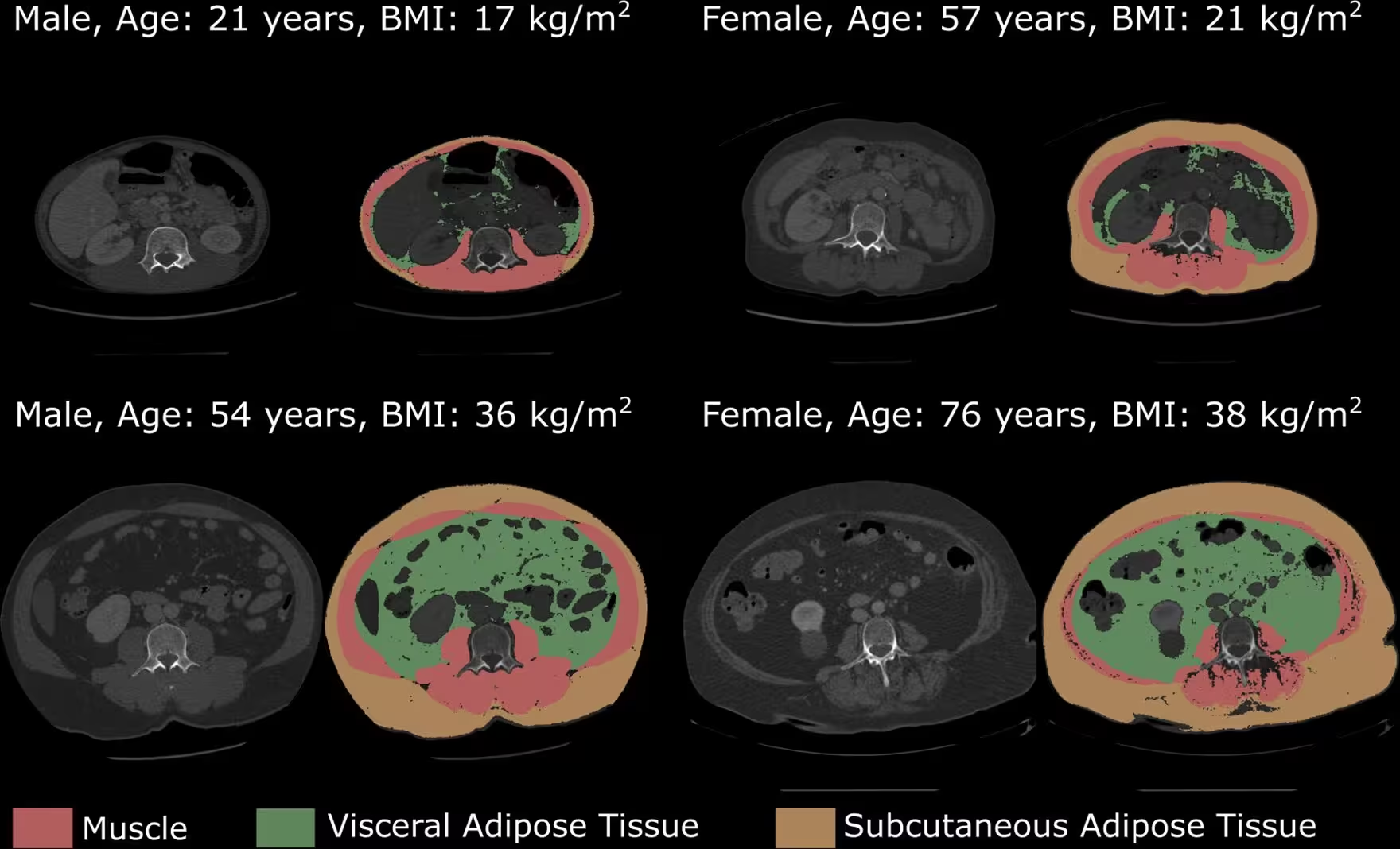
The analysis successfully confirmed these normative trajectories across both male and female cohorts. Normalized muscle cross-sectional area peaked between the ages of 30 and 39 in females and between 40 and 49 in males, after which it decreased at rates matching previous specialized pilot studies. Specifically, muscle loss accelerated in older age groups, dropping by roughly five to ten percent per decade. Meanwhile, the ratio of visceral adipose tissue, which surrounds internal organs and associates strongly with cardiometabolic diseases, to subcutaneous adipose tissue beneath the skin increased progressively with age in both sexes.
Why Body Mass Index Fails to Capture True Abdominal Fat Distribution
The massive dataset also provided a clearer picture of how body mass index relates to actual tissue distribution. The findings indicate that body mass index fails to capture an individual’s propensity to store fat viscerally rather than subcutaneously, showing only very weak correlations with the fat ratio across normal and overweight categories. However, the ratio of muscle to total abdominal adipose tissue demonstrated a moderate to strong negative correlation with body mass index, revealing that excess mass in higher weight brackets is driven primarily by fat accumulation.
When investigating low back pain, the team tested three specific hypotheses regarding muscle mass and fat distribution. Surprisingly, the statistical models revealed no significant main effect or difference in normalized muscle cross-sectional area between back pain patients and matched controls. This unexpected negative finding remained stable even after performing sensitivity analyses that adjusted for smoking history, diabetes diagnoses, prior lumbar spine surgery, and specific pain subtypes such as radiculopathy.
What the Ratio of Muscle to Total Fat Reveals About Back Pain
Although bulk skeletal muscle size did not differ significantly, the researchers discovered a subtle but statistically significant difference in the ratio of muscle to total abdominal adipose tissue. Patients diagnosed with low back pain exhibited a lower muscle-to-fat ratio than matched controls, though the effect size was very small. This suggests that the relative balance between active muscle tissue and passive fat storage might play a more important role in spinal health than overall muscle bulk alone.
The authors noted several limitations in their work, including the retrospective nature of the electronic health records and the reliance on billing diagnosis codes rather than direct physical measurements of pain severity or physical activity. Furthermore, because computed tomography scans evaluate bulk tissue rather than individual muscles or muscle quality, localized deficits in specific paraspinal muscles could not be detected. The team concludes that future digital health research should focus on regional muscle groups and more comprehensive compositional biomarkers to fully understand the mechanical drivers of back discomfort.
The research was published in PLOS Digital Health on November 19, 2026.
This article has been fact checked for accuracy, with information verified against reputable sources. Learn more about us and our editorial process.
Last reviewed on .
Article history
- Latest version
Reference(s)
- Marusich, Kathryn R.., et al. “Deep learning reveals relationships between muscle and adipose tissue and their associations with age, body mass index, and low back pain: A cross-sectional study in 10,835 subjects.” PLOS Digital Health, vol. 5, no. 11, November 19, 2026, pp. e0001788 Public Library of Science (PLoS), doi: 10.1371/journal.pdig.0001788. <https://doi.org/10.1371/journal.pdig.0001788>.
Cite this page:
- Posted by Hassan Raza