Common Painkiller Alters Brain Fluid Clearance Weeks After Mild Injury
Biology

Common Painkiller Alters Brain Fluid Clearance Weeks After Mild Injury

A single dose of buprenorphine following diffuse brain trauma shifts water channel abundance and slows waste removal in the rat thalamus.

By Hassan Raza
Published:
Email this Article
Common Painkiller Alters Brain Fluid Clearance Weeks After Mild Injury
Photo by Vitaly Gariev on Unsplash

A single dose of a common painkiller given after a mild traumatic brain injury alters key protein levels and fluid clearance in the rat brain weeks later, new research reveals. The findings highlight how post-injury medication can quietly reshape neurological recovery in region-specific ways.

How Brain Proteins Respond to Injury and Analgesics

Traumatic brain injury affects millions of people globally each year, with roughly 80 percent of cases classified as mild. Even without major visible structural damage, these mild injuries can trigger widespread cellular stress, persistent inflammation, and lasting cognitive challenges. Astrocytes, which are star-shaped glial cells responsible for maintaining brain fluid balance, blood-brain barrier integrity, and scar formation, play a central role in this delayed response.

To investigate how clinical interventions influence these cellular repairs, researchers examined the effects of buprenorphine, a semi-synthetic opioid commonly used for pain management in both human and veterinary medicine. The study evaluated male rats subjected to a central fluid percussion injury, which models diffuse brain trauma. The team analyzed four major astrocyte-associated proteins at one day and four weeks post-injury, publishing their findings in PLOS One.
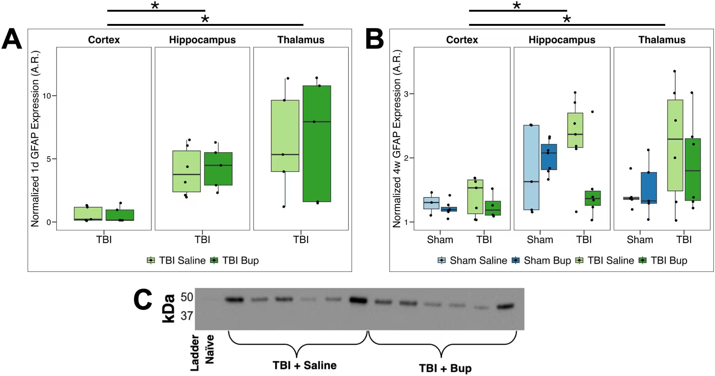
Fig 1: GFAP protein level changes associated with injury, timepoint, and buprenorphine treatment. (A) Normalized GFAP levels 1 day following cFPI-induced diffuse TBI in the right cortex, hippocampus, and thalamus. Saline-treated animals are shown in light green, and buprenorphine (bup)-treated animals are in dark green. (B) Normalized GFAP levels 4 weeks following either sham injury or cFPI-induced diffuse TBI in the right cortex, hippocampus, and thalamus. Sham/saline-treated animals are light blue, sham/bup-treated animals are dark blue, TBI/saline-treated animals are light green, and TBI/bup-treated animals are dark green. Median values and interquartile ranges are displayed for each cohort. Each dot represents individual animals. Asterisks indicate region-specific, within-injury, treatment-driven differences in GFAP expression (BH-corrected p < 0.05). (C) Representative chemiluminescent western blot with quantified band at ~50 kDa. Full uncropped blot and the corresponding total protein n
GFAP protein level changes associated with injury, timepoint, and buprenorphine treatment. (A) Normalized GFAP levels 1 day following cFPI-induced diffuse TBI in the right cortex, hippocampus, and thalamus. Saline-treated animals are shown in light green, and buprenorphine (bup)-treated animals are in dark green. (B) Normalized GFAP levels 4 weeks following either sham injury or cFPI-induced diffuse TBI in the right cortex, hippocampus, and thalamus. Sham/saline-treated animals are light blue, sham/bup-treated animals are dark blue, TBI/saline-treated animals are light green, and TBI/bup-treated animals are dark green. Median values and interquartile ranges are displayed for each cohort. Each dot represents individual animals. Asterisks indicate region-specific, within-injury, treatment-driven differences in GFAP expression (BH-corrected p < 0.05). (C) Representative chemiluminescent western blot with quantified band at ~50 kDa. Full uncropped blot and the corresponding total protein n Credit: Radina L. Lilova et al. / PLOS One

Lasting Shifts in Water Channels and Solute Clearance

The investigations focused on glial fibrillary acidic protein, a classic marker for structural remodeling, alongside three key channels regulating ion and water movement: SWELL1, TRPM4, and Aquaporin-4. In saline-treated animals, the brain showed expected increases in these markers following trauma. However, animals that received a single dose of extended-release buprenorphine fifteen minutes after the injury exhibited distinct molecular profiles weeks later.

By four weeks post-injury, buprenorphine-treated subjects showed significantly reduced levels of Aquaporin-4 in the hippocampus and thalamus compared to saline-treated injured rats. Aquaporin-4 is a critical water channel concentrated in astrocytic endfeet that helps manage fluid flow and waste clearance through the glymphatic system. To determine if these molecular shifts translated to functional changes, the researchers tracked the movement of fluorescently tagged dextran tracers infused into the brain ventricles.

The analysis revealed greater retention of the tracer within the thalamus of buprenorphine-treated injured rats compared to their saline-treated counterparts. This accumulation points to altered fluid dynamics and slower solute clearance in that specific brain region, suggesting that acute opioid exposure may influence long-term waste removal processes following head trauma.
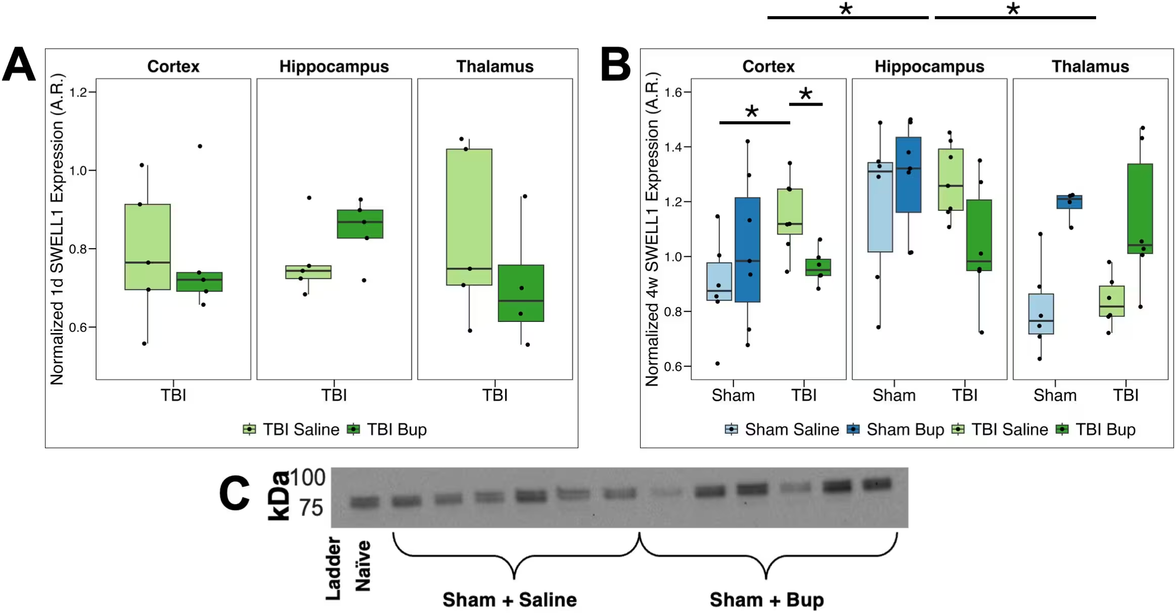
Fig 2: SWELL1 protein level changes associated with injury, timepoint, and buprenorphine administration. (A) Normalized SWELL1 expression 1 day following cFPI-induced diffuse TBI in the right cortex, hippocampus, and thalamus. Saline-treated animals are shown in light green and buprenorphine (bup)-treated animals in dark green. (B) Normalized SWELL1 expression 4 weeks following either sham injury or cFPI-induced diffuse TBI in the right cortex, hippocampus, and thalamus. Sham/saline-treated animals are light blue, sham/bup-treated animals are dark blue, TBI/saline-treated animals are light green, and TBI/bup-treated animals are dark green. Median values and interquartile ranges are displayed for each cohort. Each dot represents individual animals. Asterisks indicate region-specific, within-injury, treatment-driven differences in SWELL1 expression (BH-corrected p < 0.05). (C) Representative chemiluminescent western blot with quantified doublet band at ~85–105 kDa. Full uncropped blot and the c
SWELL1 protein level changes associated with injury, timepoint, and buprenorphine administration. (A) Normalized SWELL1 expression 1 day following cFPI-induced diffuse TBI in the right cortex, hippocampus, and thalamus. Saline-treated animals are shown in light green and buprenorphine (bup)-treated animals in dark green. (B) Normalized SWELL1 expression 4 weeks following either sham injury or cFPI-induced diffuse TBI in the right cortex, hippocampus, and thalamus. Sham/saline-treated animals are light blue, sham/bup-treated animals are dark blue, TBI/saline-treated animals are light green, and TBI/bup-treated animals are dark green. Median values and interquartile ranges are displayed for each cohort. Each dot represents individual animals. Asterisks indicate region-specific, within-injury, treatment-driven differences in SWELL1 expression (BH-corrected p < 0.05). (C) Representative chemiluminescent western blot with quantified doublet band at ~85–105 kDa. Full uncropped blot and the c Credit: Radina L. Lilova et al. / PLOS One

Interpreting Preclinical Findings for Future Care

The study underscores the complexity of treating post-traumatic pain while monitoring secondary injury mechanisms. Because virtually all standard analgesics interact with neural and glial pathways to some degree, distinguishing the physiological impacts of pain from the pharmacological effects of the medication remains a major challenge in experimental neuroscience.

The authors note several limitations to the work, including the use of only male subjects, a single post-injury dosage, and whole-tissue homogenization that could not isolate specific cellular subtypes or individual thalamic nuclei. Future research incorporating female subjects, diverse dosages, and cell-specific tracking will be essential to fully understand how analgesic choices shape neurological outcomes and how clinicians might optimize recovery strategies for human patients.

The research was published in PLOS One on November 16, 2026.

Fact Checked

This article has been fact checked for accuracy, with information verified against reputable sources. Learn more about us and our editorial process.

Last reviewed on .

Article history

  • Latest version

Reference(s)

  1. Lilova, Radina L.., et al. “Astrocyte-associated water channel changes following buprenorphine administration in a rat model of diffuse traumatic brain injury.” PLOS One, vol. 21, no. 11, November 16, 2026, pp. e0362739 Public Library of Science (PLoS), doi: 10.1371/journal.pone.0362739. <https://doi.org/10.1371/journal.pone.0362739>.

Cite this page:

Raza, Hassan. “Common Painkiller Alters Brain Fluid Clearance Weeks After Mild Injury.” BioScience. BioScience ISSN 2521-5760, 16 November 2026. <https://www.bioscience.com.pk/en/subject/biology/astrocyte-associated-water-channel-changes-following-buprenorphine-administration-in-a-rat-model-of-diffuse-traumatic-brain-injury>. Raza, H. (2026, November 16). “Common Painkiller Alters Brain Fluid Clearance Weeks After Mild Injury.” BioScience. ISSN 2521-5760. Retrieved November 16, 2026 from https://www.bioscience.com.pk/en/subject/biology/astrocyte-associated-water-channel-changes-following-buprenorphine-administration-in-a-rat-model-of-diffuse-traumatic-brain-injury Raza, Hassan. “Common Painkiller Alters Brain Fluid Clearance Weeks After Mild Injury.” BioScience. ISSN 2521-5760. https://www.bioscience.com.pk/en/subject/biology/astrocyte-associated-water-channel-changes-following-buprenorphine-administration-in-a-rat-model-of-diffuse-traumatic-brain-injury (accessed November 16, 2026).
End of the article