Nanoparticle Delivery System Protects Brain Cells From Metal Toxicity
Encapsulating a traditional herbal extract in tiny fat spheres significantly boosts its ability to defend brain tissue against manganese-induced neurodegeneration.
Manganese accumulation in the brain can destroy critical nerve centers and trigger Parkinsonian symptoms, but a plant-derived antioxidant shows surprising potential to reverse this damage when packaged in microscopic fat bubbles. Excess exposure to the essential mineral, often occurring through mining or welding work, allows metal ions to cross the blood-brain barrier and target the basal ganglia, leading to severe motor and cognitive impairments. While natural compounds like baicalein, a flavonoid extracted from Chinese skullcap roots, can fight inflammation and clear out harmful cellular waste, their poor solubility in water severely limits their effectiveness inside the body.
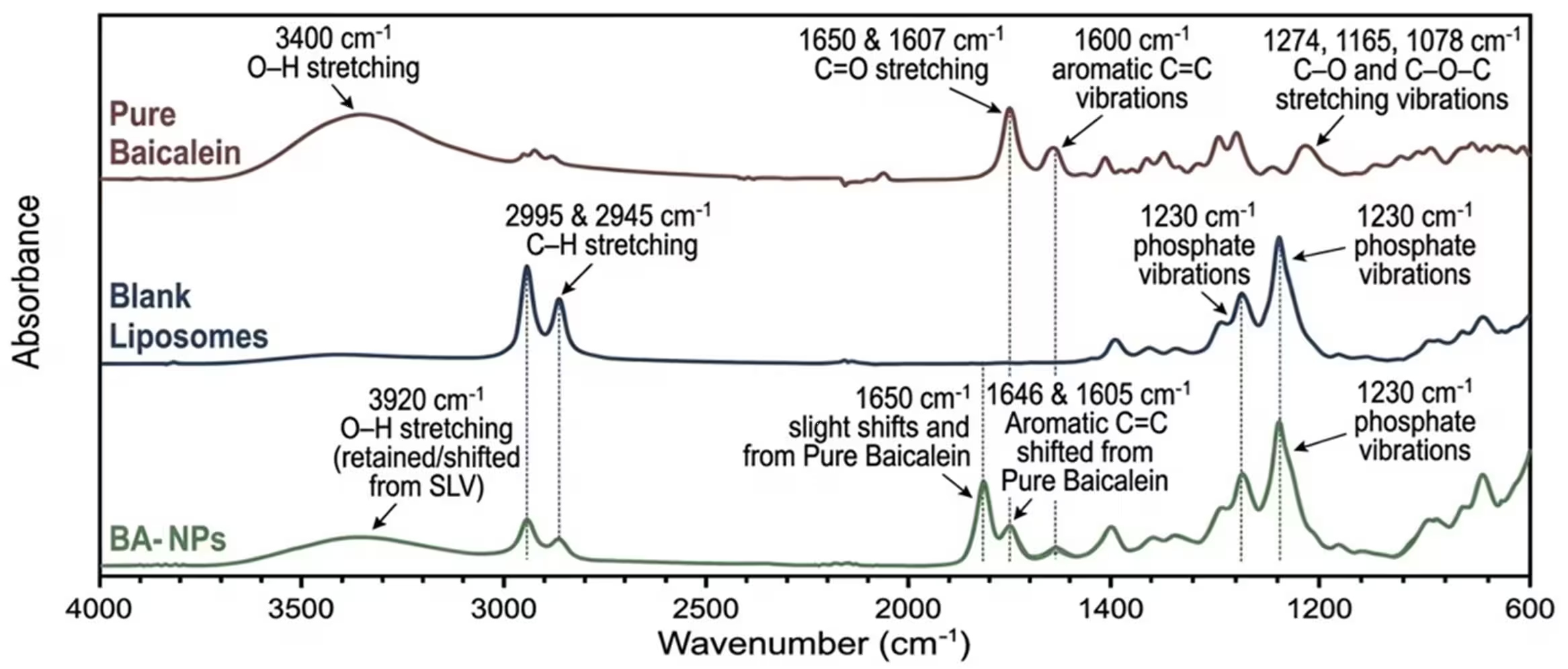
To overcome this hurdle, researchers encapsulated baicalein inside tiny lipid spheres known as liposomes, creating a delivery vehicle designed to protect the fragile compound and improve tissue absorption. The new study, published in PLOS One, tested whether these specialized nanoparticle formulations could better shield rat brains from manganese-induced neurotoxicity compared to the free chemical compound alone.
How Tiny Fat Spheres Defend Brain Cells Against Mineral Toxicity
The research team divided 60 male Wistar rats into six distinct groups to test the protective treatments over a four-week period. While one group received only a vehicle solution, others were exposed to manganese chloride at a dose of 50 milligrams per kilogram of body weight to provoke subacute neurodegeneration in the substantia nigra. Additional cohorts received free baicalein at 30 milligrams per kilogram or the liposome-encapsulated equivalent, both administered twice weekly either alone or one hour prior to manganese exposure.
Laboratory analyses of the extracted brain tissue revealed that manganese exposure severely depleted essential antioxidant defenses, including superoxide dismutase, catalase, and glutathione peroxidase enzymes. Levels of malondialdehyde, a primary biomarker for oxidative stress and cellular lipid destruction, spiked dramatically in the intoxicated animals. Furthermore, the toxic metal triggered a fourfold surge in pro-inflammatory gene expressions such as tumor necrosis factor-alpha and interleukin-6, while simultaneously crashing the production of dopamine and the vital synthesis enzyme tyrosine hydroxylase.
The Enhanced Therapeutic Power of Nanoscale Delivery
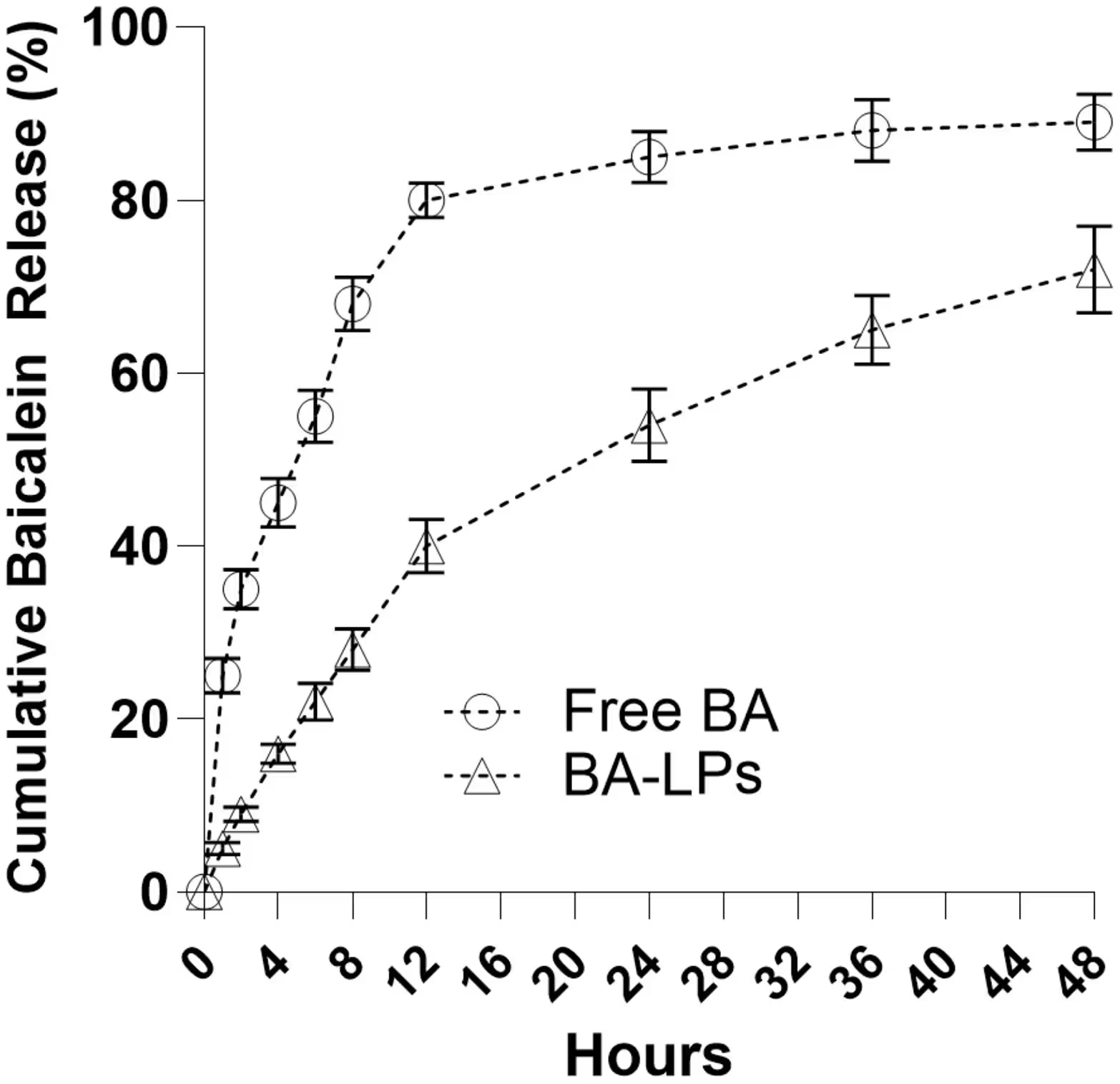
When animals were pretreated with the liposomal baicalein formulation, researchers observed a remarkable restoration of cellular health markers within the substantia nigra. The nanoparticle delivery system significantly outperformed free baicalein by restoring critical transcription factors like nuclear factor erythroid 2-related factor 2 and heme oxygenase-1 back to normal control levels. Encapsulation also slowed the release rate of the active compound in laboratory tests, extending its presence from a rapid 25-percent release in the first hour to a sustained 72-percent delivery over 48 hours.
Histological examinations confirmed that manganese-treated subjects suffered from shrunken, hyperchromatic neurons and extensive cytoplasmic vacuolation, alongside shredded mitochondrial membranes visible under transmission electron microscopy. Pretreatment with baicalein-loaded liposomes preserved normal nuclear and cellular architecture to a much higher degree, significantly reducing apoptotic cell death rates and quieting microglial and astrocytic inflammation markers.
Limitations and Future Research Directions
Despite these promising biochemical improvements, the authors noted several important constraints in their experimental design. The team did not perform pharmacokinetic or biodistribution analyses, meaning they could not directly measure how much baicalein successfully crossed the blood-brain barrier or accumulated inside specific brain tissues. Furthermore, the study lacked behavioral and functional neurological assessments to confirm whether cellular protection translated into actual motor skill recovery.
The investigators also omitted a naive untreated control group and a manganese-plus-empty-liposome group, which prevents an independent assessment of potential carrier side effects or baseline vehicle toxicity. Because the work relied solely on male rats and focused exclusively on the substantia nigra without examining other vulnerable brain regions, further preclinical trials involving dose optimization, safety profiles, and behavioral testing are required before any human clinical applications can be considered.
The research was published in PLOS One on November 16, 2026.
This article has been fact checked for accuracy, with information verified against reputable sources. Learn more about us and our editorial process.
Last reviewed on .
Article history
- Latest version
Reference(s)
- Nofal, Wesam. “Baicalein and Baicalein-loaded liposomes attenuate manganese-induced injury in the rat substantia nigra.” PLOS ONE, vol. 21, no. 11, pp. e0362801, doi: 10.1371/journal.pone.0362801. <https://doi.org/10.1371/journal.pone.0362801>.
Cite this page:
- Posted by Hassan Raza