Eight Immune Genes Linked to Severe Liver Scaring in Metabolic Disease
An integrative analysis of gut metabolite pathways and single-nucleus data reveals how immune and stellate cells interact during liver fibrosis progression.
Human liver cells show distinct shifts in immune-related genes as metabolic liver injury advances toward severe fibrosis, according to an integrative transcriptomic analysis published in PLOS One.
Connecting Gut Metabolites to Liver Fibrosis
Metabolic dysfunction-associated steatotic liver disease, or MASLD, affects millions of people globally and ranges from simple fat accumulation to severe scarring known as cirrhosis. Because routine blood tests often fail to catch advanced scarring, researchers have searched for molecular clues that better reflect the underlying damage. In the new study, investigators set out to discover whether host genes tied to gut microbial metabolites and immune responses could help distinguish early liver fibrosis from advanced disease.
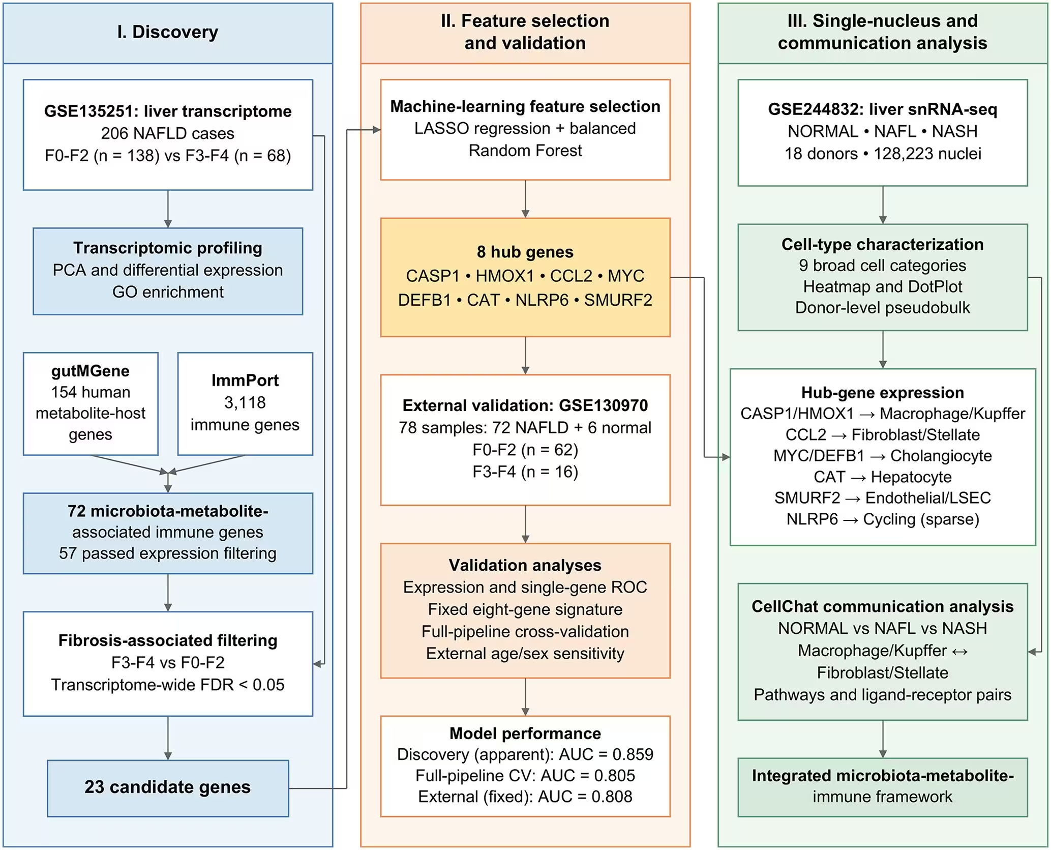
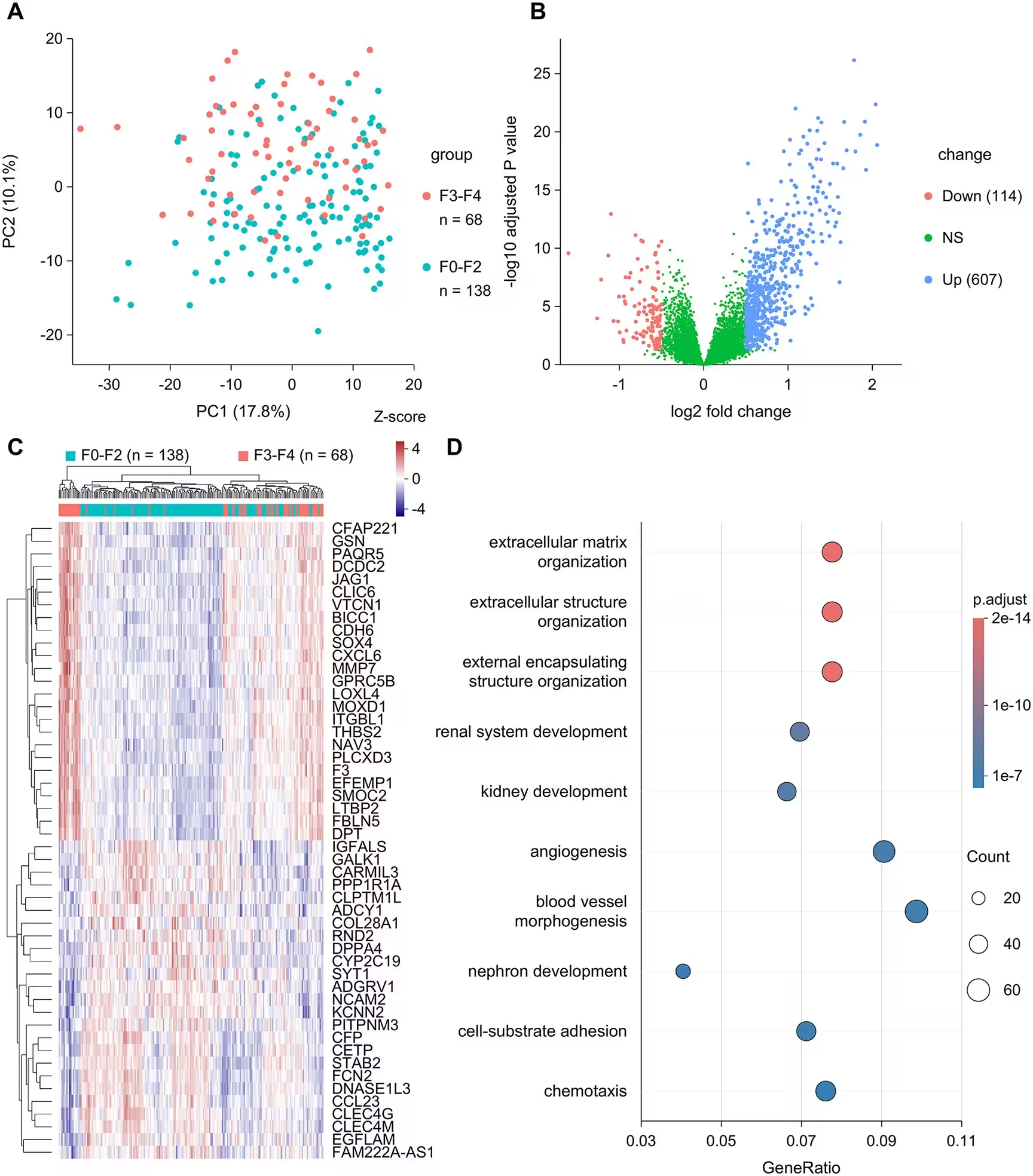
The research team began by combining two major biological databases to build a candidate list of 72 genes linked to both gut metabolites and immune functions. They then analyzed liver tissue RNA sequencing data from 206 patients with non-alcoholic fatty liver disease, comparing 138 individuals with early fibrosis against 68 patients with advanced fibrosis. Out of the 72 candidate genes, 23 showed significant differences in expression between the two groups. Using advanced machine-learning algorithms, the team narrowed this down to an eight-gene signature featuring CCL2, MYC, CASP1, CAT, DEFB1, NLRP6, HMOX1, and SMURF2.
Cellular Whereabouts of the Eight Signature Genes
To understand which liver cells produce these eight signature genes, the authors turned to single-nucleus RNA sequencing data encompassing 128,223 nuclei from 18 donors. This high-resolution mapping revealed that the genes are far from uniformly distributed. For instance, CCL2 showed its highest average expression in fibroblast and stellate cells, which are the primary drivers of scar tissue production in the liver. Meanwhile, CASP1 and HMOX1 were most prominent in macrophage and Kupffer cells, which form the liver’s frontline immune defense.
Further analysis showed that CAT was primarily expressed in hepatocytes, the main functional cells of the liver, while MYC and DEFB1 peaked in cholangiocytes, the cells lining the bile ducts. These distinct cellular locations suggest that bulk liver tissue measurements capture a complex chorus of signals coming from multiple interacting cell types rather than a single source.
Immune-Stromal Communication Across Disease States
The researchers also investigated how immune and stromal cells talk to each other across different stages of the disease using computational cell-to-cell communication tools. The analysis indicated that tissue samples from patients with non-alcoholic steatohepatitis featured a substantially larger and more active network of inferred molecular interactions than normal liver samples. Macrophages and stellate cells maintained two-way communication in all disease conditions, with signals flowing from stellate cells to macrophages making up a large share of the identified crosstalk.
While the eight-gene model demonstrated strong statistical performance when tested on the primary patient group, the authors noted several important limitations. When tested on an external mixed cohort of 78 liver biopsies, some genes did not replicate their original direction or significance, and one gene showed an opposite association. Furthermore, the database connections linking gut microbes to these host genes represent previously reported associations rather than proven causal pathways in the study participants. The investigators emphasize that the signature remains a research-stage tool that requires extensive prospective evaluation before it can guide clinical care.
The research was published in PLOS One on November 16, 2026.
This article has been fact checked for accuracy, with information verified against reputable sources. Learn more about us and our editorial process.
Last reviewed on .
Article history
- Latest version
Reference(s)
- Liu, Zhi-yun. “Integrative transcriptomic and single-nucleus analyses identify microbiota–metabolite-associated immune signatures of fibrosis severity in MASLD.” PLOS ONE, vol. 21, no. 11, pp. e0362860, doi: 10.1371/journal.pone.0362860. <https://doi.org/10.1371/journal.pone.0362860>.
Cite this page:
- Posted by Priya Sharma