New Computational Method Speeds Up Wavefront Sensing for Complex Biological Imaging
A novel approach using the discrete Fourier transform enables rapid, high-precision modeling of light scattering in thick, layered biological tissues.
High-resolution imaging of biological tissues, particularly the human retina, relies heavily on the precision of Shack-Hartmann wavefront sensors (SHWS). These devices are designed to measure the distortions of light as it passes through an optical system, allowing for the correction of aberrations that would otherwise blur the final image. While highly effective for simple, single-source light paths, these sensors encounter significant hurdles when imaging thick, scattering samples where light reflects from multiple, axially separated layers. These secondary reflections create overlapping lenslet images that bias the sensor, leading to artifactual errors in defocus and higher-order aberrations.
In a study published in Biomedical Optics Express, researchers from the Indian Institute of Science Education and Research (IISER) Berhampur have developed a powerful new computational technique to model these errors efficiently. By applying a specific coordinate transformation, the team successfully converted complex diffraction integrals into a form compatible with the discrete Fourier transform (DFT). This methodological shift overcomes the computational bottlenecks that previously limited researchers to modeling only two reflecting layers, enabling the simulation of highly complex, 75-layer retinal structures with unprecedented speed.
Overcoming the Computational Barrier
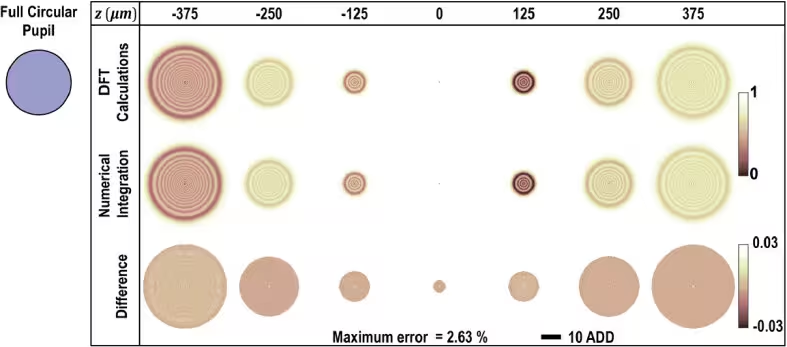
The primary challenge in modeling these sensors lies in the low Fresnel number associated with the lenslets, which traditionally complicates the use of standard Fourier-based methods. Previous approaches required direct numerical integration, a process so computationally demanding that simulating a single lenslet image in a dual-layer model could take nearly half an hour. The new DFT-based approach reduces this time by more than three orders of magnitude, making it feasible to model realistic, multilayered tissues on standard desktop hardware.
![Fig. 1: Adopted from [36]. Geometry for diffraction near the focus of a converging monochromatic spherical wavefront W at an aperture of radius a. The point P′ is specified by its position vector relative to the origin O, While Q represents a point on the wavefront W.](https://www.bioscience.com.pk/images/8697d9958e.avif)
Insights into Retinal Imaging
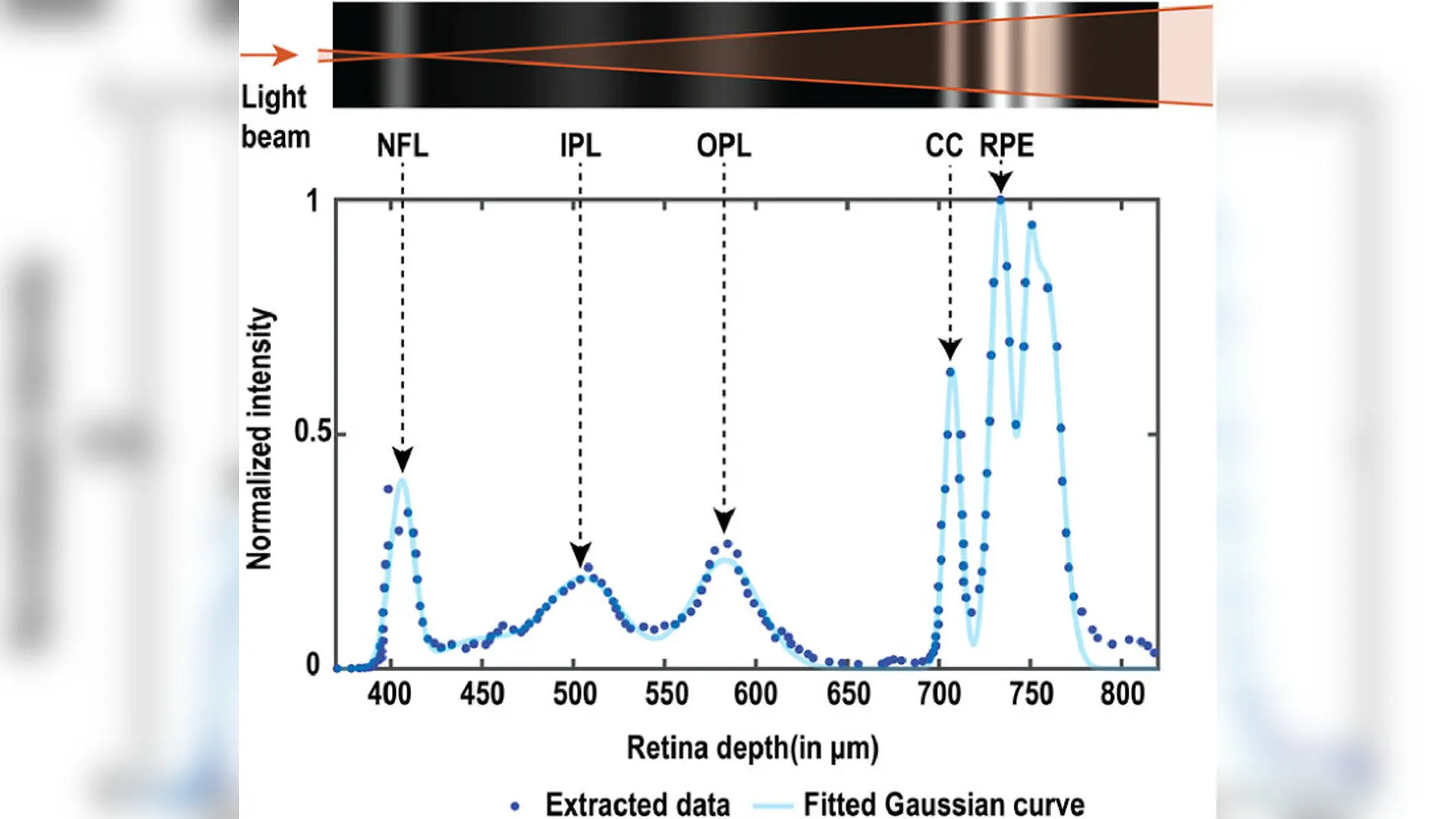
To validate their model, the researchers utilized a 75-layer retinal reflectivity profile derived from existing literature. Their simulations revealed that wavefront estimation is fundamentally sensitive to both the specific architecture of the tissue and the strategy used for centroid detection. While smaller search boxes are often used to reduce noise, the study confirms that these can inadvertently capture light from out-of-focus secondary layers, leading to significant, depth-dependent biases in the measured data.
The team found that using larger, optimized centroid search boxes significantly mitigates these errors, suppressing the abrupt variations that occur when imaging deeper layers of the retina. However, even with optimal settings, the multilayered nature of the retina introduces a residual constant defocus bias that is notably larger than what would be predicted by a simplified two-layer model. This highlights the necessity of using comprehensive, multilayered simulations to accurately interpret data from complex biological samples.
Future Implications for Adaptive Optics
The ability to rapidly and accurately simulate these artifacts has immediate implications for the development of adaptive optics in clinical settings. Beyond retinal imaging, the researchers suggest that this framework could be extended to other areas of biomedical imaging, such as the study of animal retinas or the microscopy of thick, multicellular samples. By enabling faster, more accurate predictions of wavefront sensing errors, this work provides a vital tool for researchers looking to push the boundaries of high-resolution imaging in complex, volumetric environments.
The study also points to practical mitigation strategies for current imaging systems. While dynamic beacon positioning remains a primary method for focusing on the brightest layer of interest, the researchers suggest that pairing small on-axis illumination with polarizers offers a robust, easy-to-implement alternative for minimizing corneal reflections and other artifactual aberrations. As adaptive optics technology continues to evolve, these computational advancements will play a crucial role in ensuring that the next generation of imaging systems can overcome the inherent challenges posed by the complex, layered structures of the human eye.
The research was published in Biomedical optics express on August 1, 2026.
This article has been fact checked for accuracy, with information verified against reputable sources. Learn more about us and our editorial process.
Last reviewed on .
Article history
- Latest version
Reference(s)
- Sahoo, Paresh Kumar., et al. “Evaluation of Shack-Hartmann wavefront sensing artifacts due to reflectivity variations across thick layered samples using the discrete Fourier transform.” Biomedical Optics Express, vol. 17, no. 8, July 10, 2026, pp. 4115 Optica Publishing Group, doi: 10.1364/BOE.596303. <https://doi.org/10.1364/BOE.596303>.
Cite this page:
- Posted by Aisha Ahmed