Skin Biopsies Reveal Early Parkinson’s Disease Protein Clumps
A quantitative analysis of skin tissue samples shows that misfolded proteins multiply faster in patients with specific early sleep disorder symptoms.
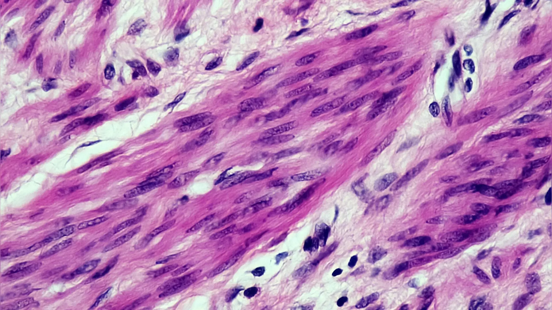
Skin biopsies can reveal signs of Parkinson’s disease before movement symptoms fully develop, according to a recent analysis of data from the Parkinson’s Precision Medicine Initiative.
Researchers examined tissue samples from 93 participants to track how abnormal protein clumps spread through peripheral nerves in people with sleep disorders and established diagnoses. The study, published in PLOS One, demonstrates that a simple skin test can detect misfolded alpha-synuclein proteins, which are a hallmark of the condition.
Detecting Protein Aggregates in Sleep Disorder Patients
Parkinson’s disease is traditionally diagnosed based on motor difficulties such as tremors and stiffness, but non-motor symptoms often appear years earlier. One of the most prominent early signs is REM sleep behavior disorder, a condition where individuals physically act out vivid dreams due to a loss of normal muscle paralysis during sleep.
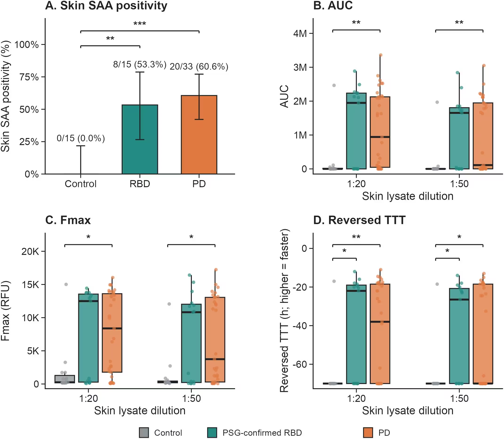
In this cross-sectional study, investigators evaluated skin samples from healthy controls, people with polysomnography-confirmed REM sleep behavior disorder, and patients with established Parkinson’s disease. Polysomnography is a comprehensive sleep study that records brain waves, eye movements, and muscle activity. The assay measures how quickly these disease-associated proteins multiply in a laboratory test.
Protein seeding was detected in 53.3 percent of participants with confirmed sleep disorders and 60.6 percent of those with established Parkinson’s disease. None of the healthy control participants showed positive results. Furthermore, the protein amplification rate was significantly faster in tissue samples from individuals with the sleep disorder compared to healthy controls.
Quantitative Kinetics Reveal Subgroup Differences
Beyond simply noting whether a test is positive or negative, the research team analyzed the speed and intensity of protein growth. These quantitative metrics, including area under the curve and maximum fluorescence measurements, provided a more detailed picture of how disease pathology varies among patients.
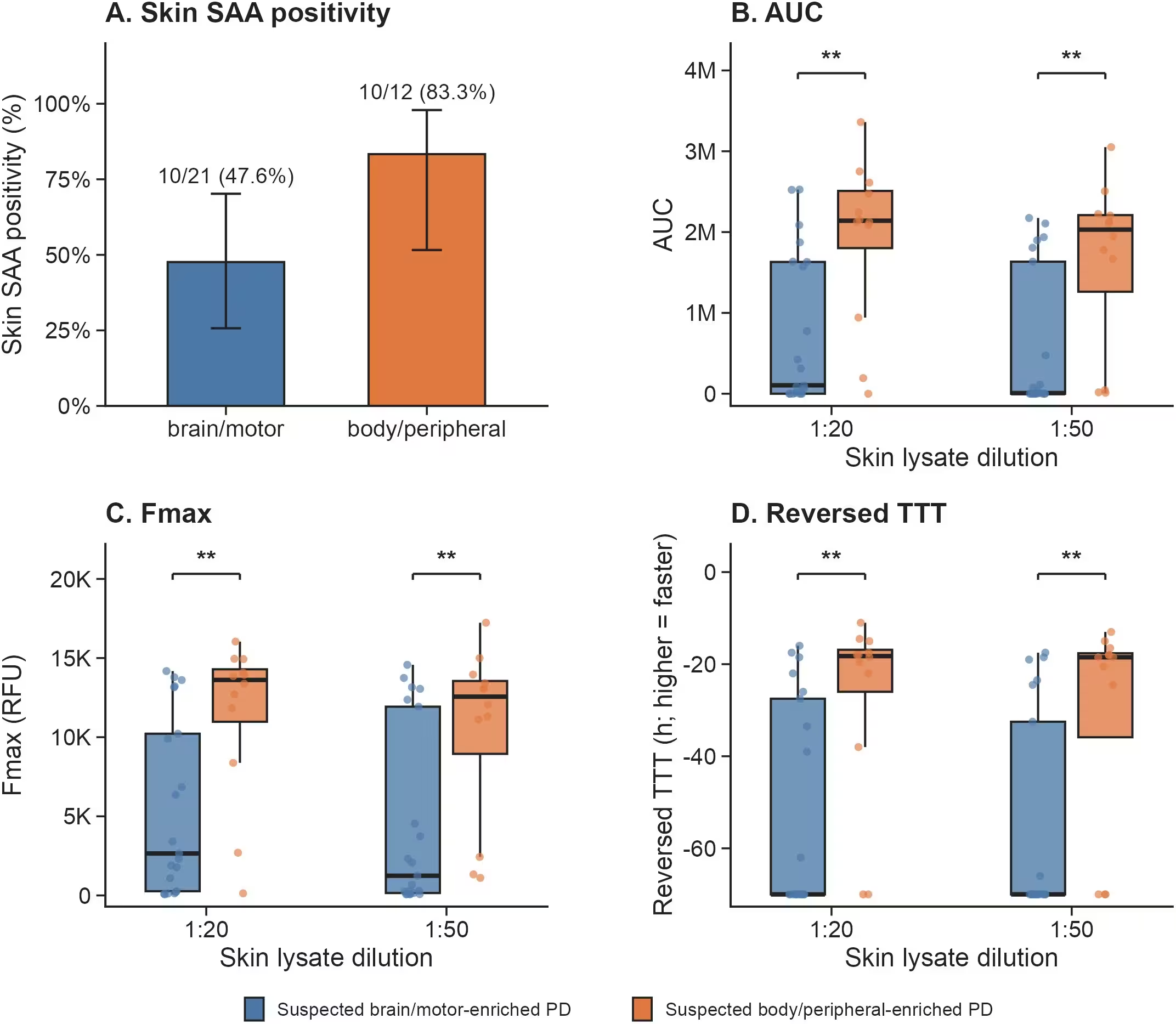
Within the Parkinson’s disease group, patients were classified into exploratory clinical phenotypes based on their symptom profiles. Individuals categorized into a suspected body-first or peripheral-enriched pathway showed markedly higher seeding kinetics than those with brain-first or motor-enriched features. Statistical models confirmed that these differences remained robust even after adjusting for participant age and alternative classification criteria.
Understanding Symptom Burden and Limitations
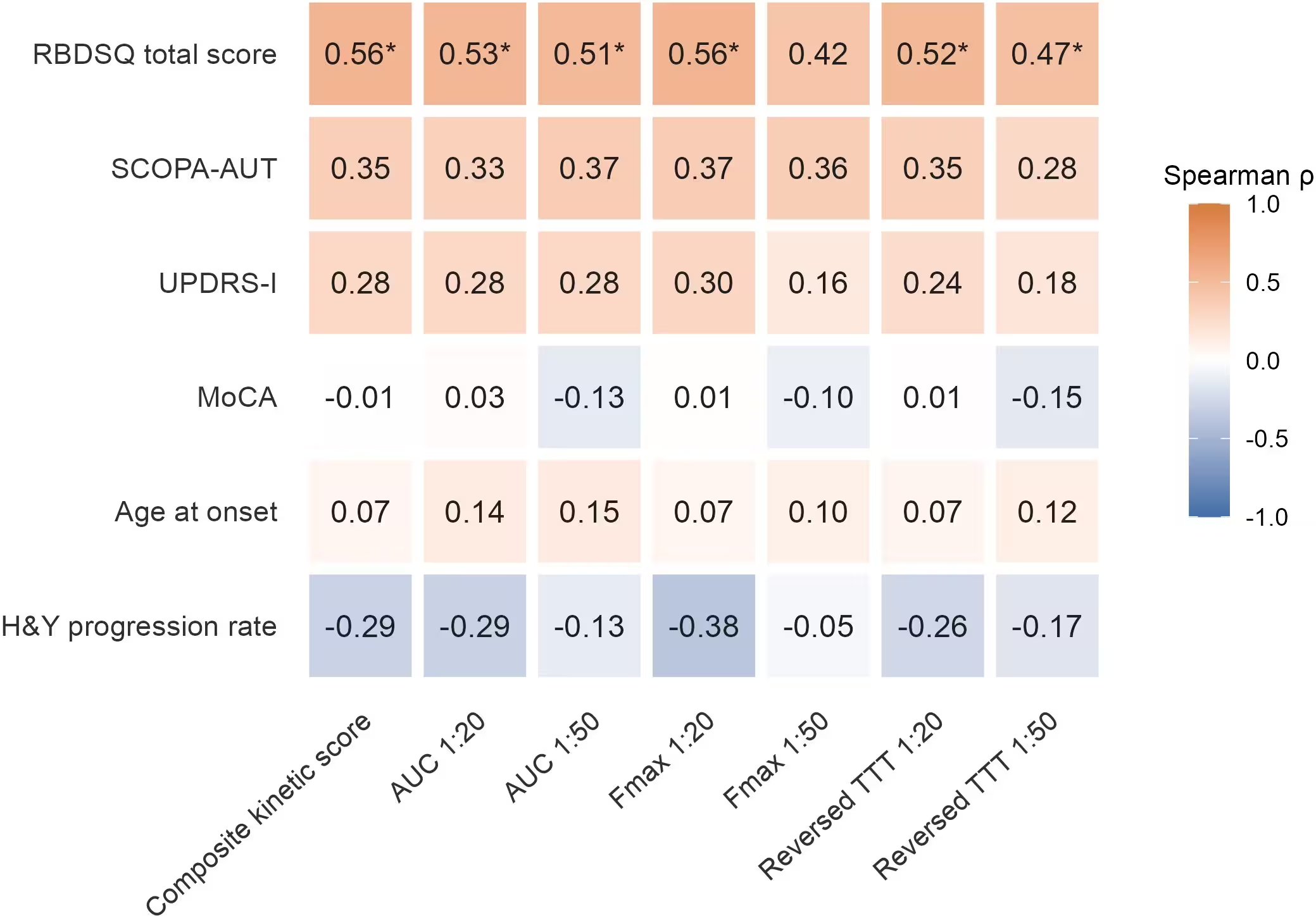
Further analysis showed a clear link between the severity of sleep disturbance scores and protein amplification speed. Patients who reported more frequent dream-enactment behaviors exhibited faster protein aggregation in their skin tissue samples.
However, the authors noted several limitations in their work. The study relied on a modest sample size, particularly within the prodromal sleep disorder group, and used a cross-sectional design that captures only a single point in time rather than tracking disease progression longitudinally. Additionally, symptom questionnaires used in established patients differ from the rigorous sleep studies used to confirm prodromal diagnoses.
These findings point to the potential usefulness of peripheral tissue tests for identifying biological subtypes before severe motor decline occurs. Future prospective studies with larger cohorts and longer follow-up periods will be necessary to determine how skin test kinetics can be applied in clinical trials or patient stratification.
The research was published in PLOS One on November 20, 2026.
This article has been fact checked for accuracy, with information verified against reputable sources. Learn more about us and our editorial process.
Last reviewed on .
Article history
- Latest version
Cite this page:
- Posted by Susan White