New AI Method Accelerates Brain Stiffness Scans By 19-Fold
A physics-informed deep learning approach successfully maps tissue elasticity from heavily undersampled data without sacrificing quantitative accuracy.

A team of researchers has developed a new physics-informed deep learning method that significantly accelerates Magnetic Resonance Elastography, a medical imaging technique used to map tissue stiffness and detect neurological disorders. By estimating shear wave speed directly from heavily undersampled data, the technique cuts scan times by up to 19-fold while maintaining high quantitative accuracy.
Magnetic Resonance Elastography, known as MRE, is a specialized quantitative imaging method that assesses the biomechanical properties of internal organs and tissues. By applying external mechanical vibrations, the technique generates shear waves that travel through tissue, causing microscopic displacements. These movements are captured by specialized magnetic resonance sequences and converted into shear wave speed maps, which help doctors detect localized changes in tissue stiffness associated with conditions like Alzheimer’s disease. However, traditional MRE requires multiple time-resolved acquisitions, resulting in long scan times that can cause patient discomfort and limit temporal resolution.
How End-to-End Deep Learning Transforms Brain MRE
To overcome these speed barriers, the authors of the study, featuring researchers from the Physikalisch-Technische Bundesanstalt and Charité – Universitätsmedizin Berlin, designed an end-to-end trainable neural network framework. The details are available in a study published in IEEE Transactions on Biomedical Engineering. Traditional pipelines separate image reconstruction from the elastography inversion calculation, meaning any errors in the initial image processing propagate through to the final stiffness map. The new approach combines model-based deep learning image reconstruction with a phase-gradient inversion method called k-MDEV, treating both steps as a unified, trainable pipeline.
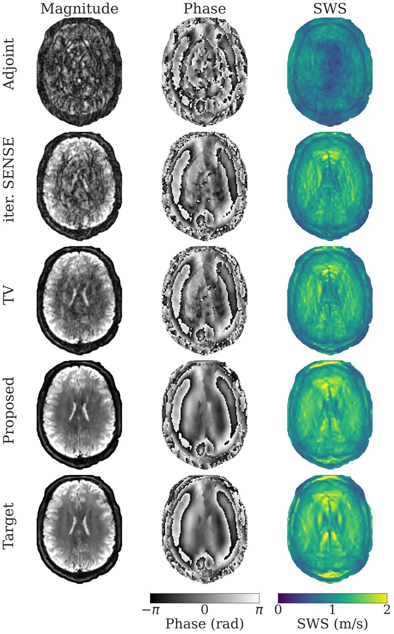
The network utilizes algorithm unrolling, alternating between convolutional neural network blocks that reduce noise and data consistency layers that anchor the estimates to the raw measurements. By training the system using both qualitative image targets and quantitative shear wave speed targets, the neural network learns a task-specific regularization tailored directly to stiffness quantification.
Evaluating Performance on Accelerated Brain Scans
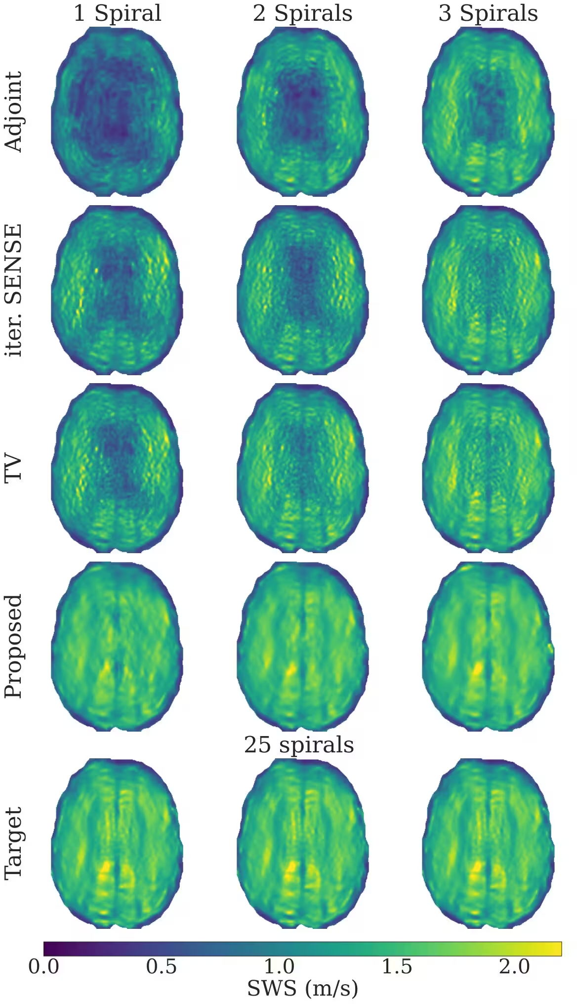
The researchers evaluated their framework using data collected from 13 healthy volunteers at 3 Tesla, alongside retrospectively undersampled Cartesian reference scans. The study simulated accelerated data acquisition using one, two, or three spiral trajectories, corresponding to undersampling factors of 19, 9.5, and 6.3, respectively. An ablation study examining the number of unrolled iterations revealed that increasing the unrolling depth from one to six steps improved shear wave speed estimation quality by 18 percent.
Quantitative metrics showed that the proposed end-to-end approach substantially outperformed conventional parallel imaging and total variation regularization methods. On the 19-fold undersampled test set, the new method achieved a structural similarity index of 0.93 and a normalized root mean squared error of 0.14 for shear wave speed maps, representing a marked improvement over traditional baseline techniques.
Potential Limitations and Future Clinical Directions
While the results highlight significant improvements in reconstruction quality and data efficiency, the authors note several limitations. The training process requires substantial computational resources, taking roughly one week of total training time on high-end graphics processing units. Furthermore, the current evaluation was conducted exclusively on healthy volunteers, meaning that systematic clinical validation in larger patient cohorts with pathological stiffness abnormalities remains an essential next step.
Despite these challenges, the ability to generate reliable stiffness maps from single-shot spiral acquisitions opens the door to real-time mechanical mapping. This acceleration could facilitate free-breathing acquisitions and improve clinical workflows for challenging examinations such as abdominal or pediatric imaging.
The study was posted to the arXiv preprint server on November 19, 2026.
This article has been fact checked for accuracy, with information verified against reputable sources. Learn more about us and our editorial process.
Last reviewed on .
Article history
- Latest version
Reference(s)
- Martin, Stefan., et al. “Physics-Informed Deep Learning for Shear Wave Speed Estimation in MR Elastography.” IEEE Transactions on Biomedical Engineering, January 1, 2026, pp. 1-12. Institute of Electrical and Electronics Engineers (IEEE), doi: 10.1109/TBME.2026.3666306. <https://doi.org/10.1109/TBME.2026.3666306>.
Cite this page:
- Posted by Susan White