Hidden Markov Model Reveals How Brain Dynamics Shift Before Seizures
Mathematical modeling of intracranial recordings from 30 patients improves the detection of seizure-generating brain tissue.
Brain signals in patients with drug-resistant epilepsy undergo subtle shifts long before a seizure officially begins, according to a mathematical modeling study. Researchers examined intracranial recordings from 30 patients and found that tracking how brain electrical patterns transition between hidden states can significantly improve the accuracy of locating seizure-generating tissue.
How Hidden States Map Brain Activity
For individuals with drug-resistant epilepsy who do not respond to medications, surgical removal of the epileptogenic zone, which is the specific brain region responsible for triggering seizures, offers the highest chance of long-term seizure freedom. To pinpoint this zone before surgery, medical teams often rely on stereo-electroencephalography, a procedure where multiple needle electrodes are surgically implanted deep into the brain. These sensors produce complex, multi-dimensional data streams consisting of hundreds of individual channels that record electrical activity over time.
Traditionally, clinicians inspect these signals visually or apply automated metrics that evaluate each channel in isolation during specific time windows. However, seizures are fundamentally network phenomena involving coordinated shifts across multiple brain regions. To capture these dynamics better, the study published on the arXiv preprint server introduced a joint Hidden Markov Model framework. In this mathematical approach, a hidden probabilistic structure describes how each channel moves between underlying states.
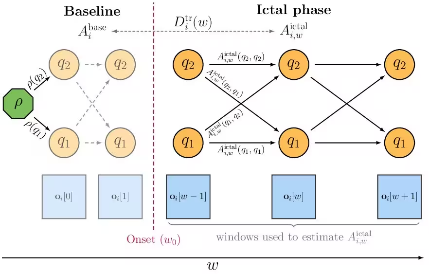
The authors, including researchers from the University of Padova and the IRCCS Institute of Neurological Sciences of Bologna, designed the model so that all channels share the same set of hidden states and emission characteristics, while each individual channel maintains its own unique transition probability matrix. This structure allows the algorithm to evaluate how easily a specific region shifts from a normal baseline state toward abnormal, seizure-like dynamics as a seizure unfolds.
Measuring Divergence Between Baseline and Seizure Onset
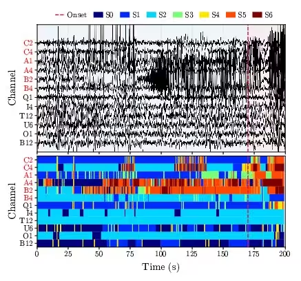
To quantify these behavioral changes, the research team calculated the Kullback-Leibler divergence, a statistical measure of how one probability distribution differs from another, comparing a channel’s transition probabilities during the inter-ictal period (the resting time between seizures) against the moment of seizure onset. They analyzed 200-second epochs from 30 patients, encompassing a 170-second pre-onset baseline and the first 30 seconds of the ictal phase. Clinically validated thermocoagulated channels, which represent the actual surgical targets treated with radiofrequency ablation, served as the ground truth for evaluating performance.
When tested across the patient cohort, the joint model demonstrated superior performance compared to a standard benchmark approach that only looked at static emission distributions without modeling temporal transitions. Specifically, the top-performing variant achieved a mean area under the receiver operating characteristic curve of 0.797, successfully outperforming the benchmark in 27 out of the 30 patients. Meanwhile, the benchmark method performed worse than a random classifier in six of those patients when restricted to basic energy features, highlighting the necessity of capturing temporal evolution.
Limitations and Future Clinical Directions
Despite these promising results, the study authors noted several technical limitations that should be addressed in future work. The current model assumes that features within the same frequency band are conditionally independent, a simplification that helps computational tractability but may not fully reflect complex physiological correlations in neural data. Furthermore, the framework was evaluated retrospectively on a specific dataset, meaning prospective validation across larger, multi-center cohorts will be required before clinical translation.
By moving beyond static snapshots and explicitly modeling the temporal transitions of brain signals, this data-driven framework offers a robust way to analyze intracranial recordings. The findings point toward better computational tools that could eventually assist neurologists in interpreting complex stereo-electroencephalography data and planning safer, more precise surgical interventions for epilepsy.
The study was posted to the arXiv preprint server on November 19, 2026.
This article has been fact checked for accuracy, with information verified against reputable sources. Learn more about us and our editorial process.
Last reviewed on .
Article history
- Latest version
Reference(s)
- Tacconi, Giulia. “Transition Dynamics Matter: Hidden Markov Scoring of Epileptogenic Channels in SEEG.” arXiv.org <http://arxiv.org/abs/2611.26142v1>.
Cite this page:
- Posted by Tariq Hassan