New Microneedle Patch Detects Early Kidney Disease Without The Need For Blood Draws
Researchers are developing a minimally invasive diagnostic method that could detect kidney disease much earlier, potentially improving patient outcomes.
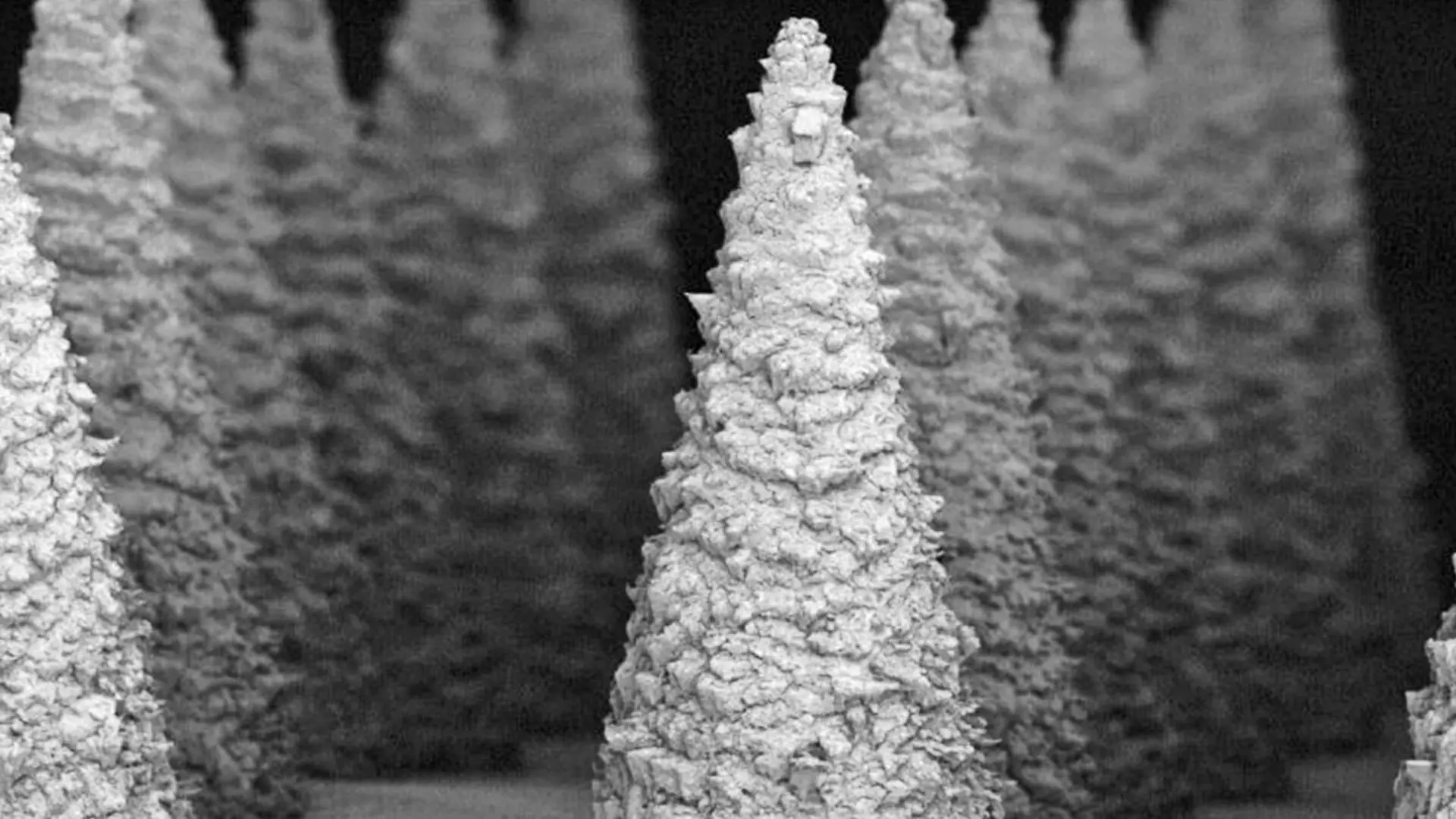
A team of researchers has unveiled a new, minimally invasive diagnostic tool capable of identifying kidney injury long before physical symptoms appear. By utilizing a specialized microneedle patch, the scientists have demonstrated a way to sample interstitial fluid through the skin, offering a potential breakthrough for early clinical intervention in kidney health.
The findings, published in the journal Advanced Materials, detail a system designed to overcome the traditional barriers of biomarker monitoring. Kidney disease is notoriously difficult to diagnose in its nascent stages, often remaining asymptomatic until the damage is significant. The new device aims to change this by providing a reliable, user-friendly method for tracking biological markers outside of a traditional hospital setting.
Advanced Material Innovation
At the core of the technology is a microneedle array coated in a metal-organic framework (MOF). This porous shell serves a dual purpose: it acts as a capture mechanism for specific biomarkers and functions as a protective shield for the sensitive antibodies trapped within. Specifically, the patch is engineered to detect neutrophil gelatinase-associated lipocalin (NGAL), a protein that acts as an early warning signal for acute kidney injury.
One of the most significant hurdles in diagnostic testing is the requirement for cold-chain logistics, which necessitates that samples and testing kits be kept refrigerated to maintain chemical stability. The research team, led by Srikanth Singamaneni of the department of mechanical engineering and materials science at Washington University in St. Louis, successfully demonstrated that their MOF-coated needles can preserve the biological function of antibodies for up to four weeks at temperatures as high as 50 C (122 F).
Transforming Point-of-Care Diagnostics
Current clinical protocols for measuring NGAL levels typically require a blood draw, which is invasive and necessitates professional laboratory analysis. By moving the detection process to the skin’s interstitial fluid via a simple patch, the researchers hope to facilitate remote monitoring and point-of-care diagnostics that do not require refrigeration or specialized medical staff.
“This metal-organic framework encapsulation is a simple and highly effective way to create microneedle sensors that are resilient to environmental challenges and provide a scalable path to minimally invasive biosensing for at-home or remote health monitoring,” Singamaneni noted regarding the potential impact of the technology.
The study marks a significant evolution in biosensor development, as it is the first to prove that biomolecules can remain functional while encapsulated on microneedles in this manner. The team, which included doctoral student Yixuan Wang and collaborators from WashU Medicine and Texas A&M University, built upon previous work involving microneedle technology to achieve the necessary sensitivity for clinical utility.
The research received support from the National Science Foundation, the National Institutes of Health, the Congressionally Directed Medical Research Programs, and the VA Merit program. Additionally, the Office of Technology Management at Washington University in St. Louis has licensed the underlying plasmonic-fluor technology, which was developed by Singamaneni and Jeremiah J. Morrissey.
This article has been fact checked for accuracy, with information verified against reputable sources. Learn more about us and our editorial process.
Last reviewed on .
Article history
- Latest version
Cite this page:
- Posted by David Anderson