Blood Protein Map in Sickle Cell Patients Reveals 58 Novel Genetic Targets
Proteogenomic analysis of 343 patients identifies key plasma markers and potential regulators of fetal hemoglobin production.
A sweeping genetic analysis of blood proteins in sickle cell disease patients has uncovered 560 genetic control points that shape the human plasma proteome, revealing unexpected biological clues about the inherited blood disorder. Sickle cell disease is a severe monogenic condition affecting over 7 million people worldwide, caused by mutations in the beta-globin gene that deform red blood cells into rigid sickle shapes. While therapies remain scarce and expensive, researchers have struggled to identify new drug targets tailored specifically to this underserved patient population.
Pinpointing Protein Control Points in 343 Sickle Cell Patients
Led by scientists aiming to map how genetic variants alter blood protein levels, the team analyzed plasma samples from 343 adult participants of African ancestry with the severe HbSS genotype from the GEN-MOD cohort. None of the participants had received a blood transfusion or hydroxyurea treatment in the 90 days before sample collection. Using an advanced antibody-based proteomic platform capable of scanning thousands of circulating molecules, the authors measured 5,411 distinct plasma proteins and tested their links against approximately 4.6 million imputed genetic variants.
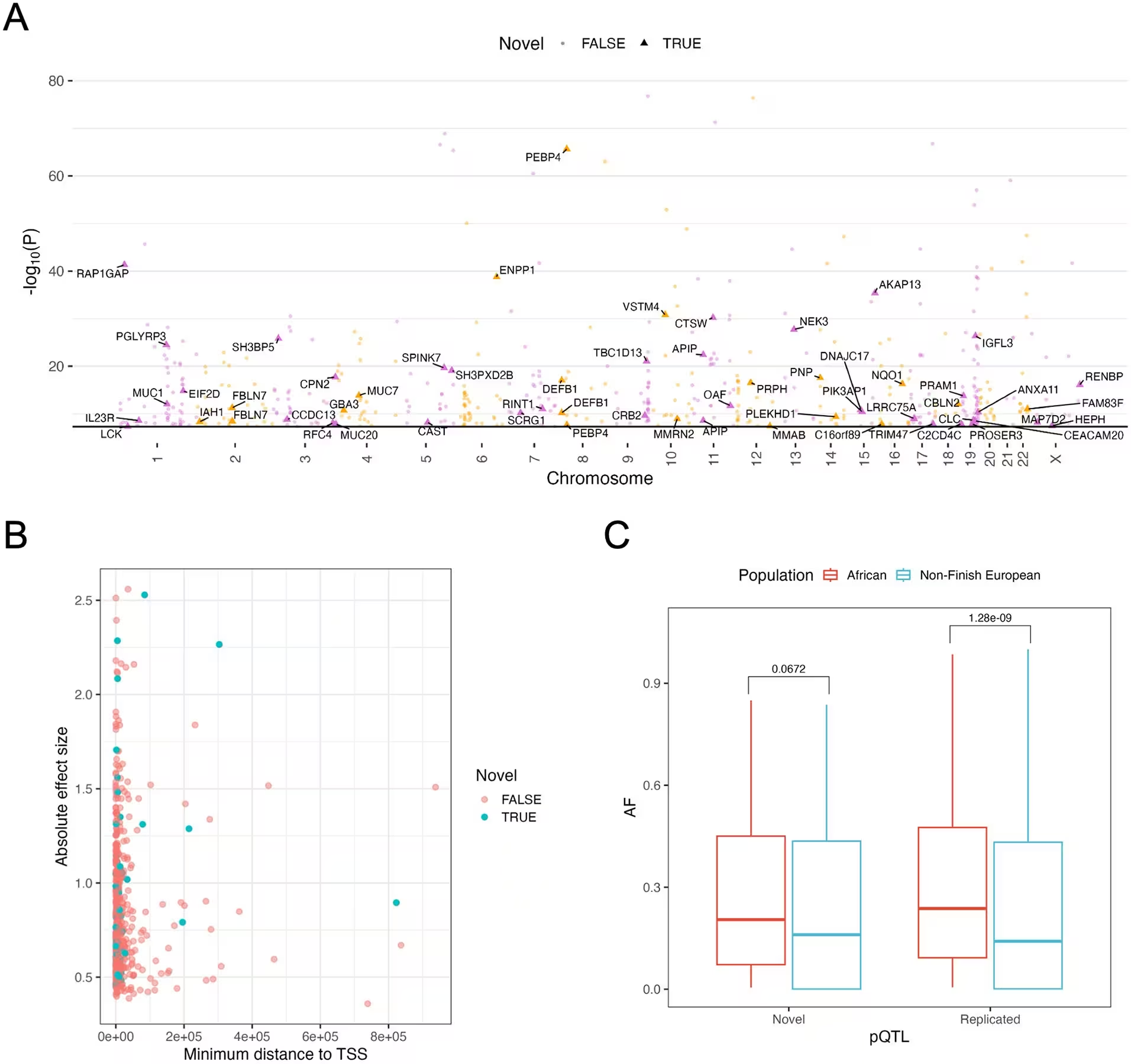
After rigorous statistical filtering and conditional analyses published in PLOS Genetics, the investigators identified 529 cis-protein quantitative trait loci and 31 trans-protein quantitative trait loci, which together implicate 467 unique proteins. Crucially, 58 of these genetic associations, representing 10 percent of the discoveries, were entirely novel. Most of these newly uncovered variant-protein pairs involved proteins that were missing from older proteomic assays or represented signals that did not overlap with previous population databases, pointing to potential sickle cell-specific biological mechanisms.
Comparing Genetic Effects Across Healthy and Affected Cohorts
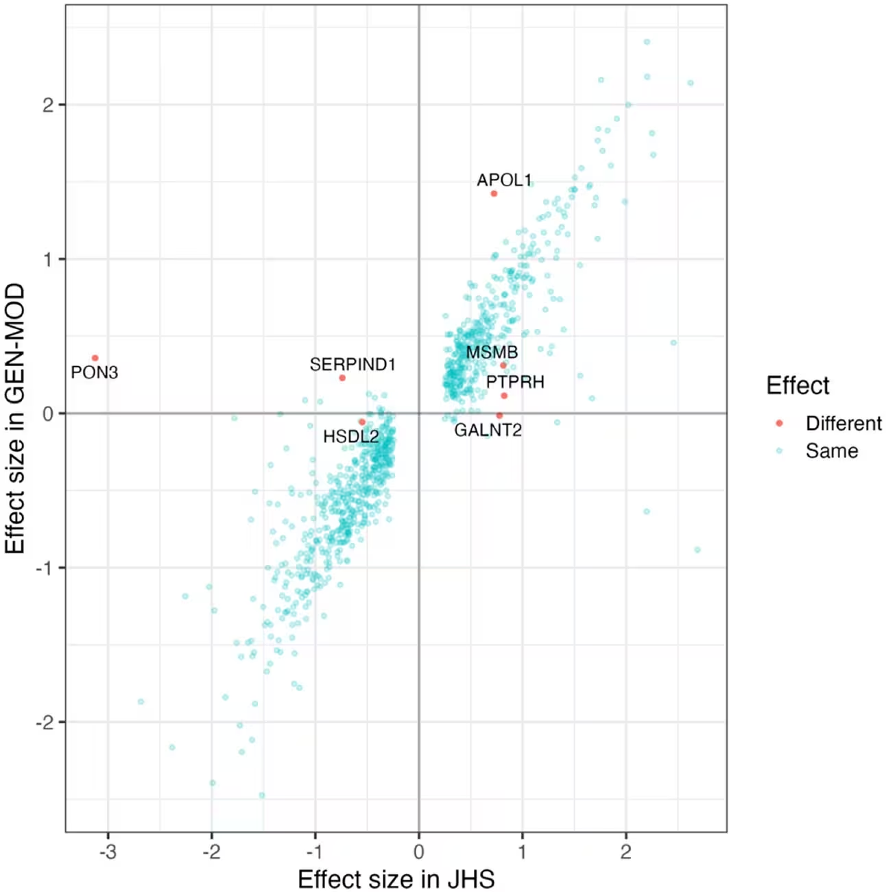
To determine whether standard population data applies to diseased individuals, the researchers compared their findings with genetic and proteomic maps from the Jackson Heart Study, a large population-based cohort of Black adults. The overall effect sizes of the protein quantitative trait loci showed strong consistency between the two groups, with a correlation coefficient of 0.90. This indicates that the core genetic architecture regulating most circulating proteins remains largely undisturbed by the sickle cell genotype.
However, the team also spotted seven notable exceptions where genetic effect sizes diverged significantly between healthy and sickle cell participants. One prominent example involved the haptoglobin gene region, where constant red blood cell destruction and cell-free hemoglobin scavenging in sickle cell patients appear to mask typical genetic associations. Another critical divergence appeared at the apolipoprotein L1 gene, where risk variants linked to kidney disease showed drastically amplified plasma protein signatures in sickle cell carriers.
Unmasking Candidate Regulators of Fetal Hemoglobin Production
Because fetal hemoglobin is a powerful natural buffer that protects patients from painful crises, stroke, and early death, finding ways to boost its synthesis is a major clinical priority. By integrating their proteomic quantitative trait loci with a large meta-analysis of fetal hemoglobin genome-wide association studies, the team applied two-sample Mendelian randomization to test whether specific plasma proteins causally influence fetal hemoglobin levels.
The causal inference analyses flagged several candidate proteins, highlighting pentraxin 3 as a marker of inflammation associated with increased fetal hemoglobin. They also identified ectonucleotide pyrophosphatase phosphodiesterase family member 5, an enzyme involved in nucleotide metabolism, as a putative novel inducer of fetal hemoglobin production. Conversely, genetically controlled levels of the zeta subunit of embryonic hemoglobin showed unexpected negative associations with fetal hemoglobin output. While these statistical models offer promising therapeutic leads, the authors emphasize that further experimental validation in cellular and animal models will be necessary before these targets can translate into human therapies.
The research was published in PLOS Genetics on November 16, 2026.
This article has been fact checked for accuracy, with information verified against reputable sources. Learn more about us and our editorial process.
Last reviewed on .
Article history
- Latest version
Reference(s)
- Groza, Cristian., et al. “Proteogenomic analysis of 5,411 plasma proteins in sickle cell disease patients.” PLOS Genetics, vol. 22, no. 11, November 16, 2026, pp. e1012377 Public Library of Science (PLoS), doi: 10.1371/journal.pgen.1012377. <https://doi.org/10.1371/journal.pgen.1012377>.
Cite this page:
- Posted by Elizabeth Taylor