Hidden Chromosome Deletions Drive Severe Androgen Insensitivity Syndrome
Genetics

Hidden Chromosome Deletions Drive Severe Androgen Insensitivity Syndrome

A new genetic analysis reveals why large structural variants in the X chromosome are five times more common in patients with androgen insensitivity.

By Elizabeth Taylor
Published:
Email this Article
Hidden Chromosome Deletions Drive Severe Androgen Insensitivity Syndrome
Photo by Warren Umoh on Unsplash

A 24-year-old patient carrying a large genetic deletion on their X chromosome has provided the first direct molecular evidence of how microhomology-dependent repair mechanisms can permanently disrupt the androgen receptor gene. Researchers analyzed a 46,XY individual with complete androgen insensitivity syndrome, a condition where the body cannot respond properly to male sex hormones despite having typical male chromosomes.

The patient presented during adolescence with primary amenorrhea and a history of an inguinal hernia treated in early childhood. Clinical hormone evaluations revealed elevated total testosterone and increased luteinizing hormone levels alongside normal follicle-stimulating hormone. Pelvic magnetic resonance imaging confirmed the absence of a uterus, cervix, and upper vagina, paired with bilateral gonadal-like structures located in the iliac fossae. Cytogenetic testing confirmed a 46,XY karyotype.

How Exome Sequencing and High-Resolution Mapping Uncovered the Deletion

Initial whole-exome sequencing pointed toward a genomic loss on chromosome Xq12. To pinpoint the boundaries of this structural alteration, the team utilized multiplex ligation-dependent probe amplification alongside polymerase chain reaction and Sanger sequencing. These combined approaches, published in Human Mutation, revealed a massive deletion spanning exons 6 through 8 of the androgen receptor gene. This specific region encodes the ligand-binding domain, which is the functional portion of the receptor responsible for hormone binding.
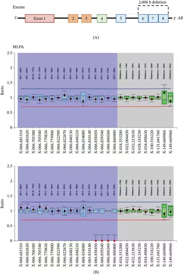
Figure 1: Identification and refinement of deletions in the AR gene using exome sequencing, MLPA (multiplex ligation‐dependent probe amplification), and Sanger sequencing. (A) Schematic representation of the AR gene exons (1–8), with a 2008‐bp deletion initially identified by exome sequencing, spanning Exons 6–8. (B) MLPA analysis identified a deletion encompassing Exons 6–8 of the AR gene. Probe signal intensities from the patient were compared with reference samples to assess copy number variation. The blue and red horizontal lines represent arbitrary thresholds set at approximately ±0.3 from the average probe value across reference samples. Blue and green boxes correspond to the 95% confidence intervals (CIs) for each probe in the reference group. Probes in the blue‐shaded zone target exonic regions of interest. Probes in the gray‐shaded zone serve as internal controls. Black dots represent patient probe ratios within the 95% CI; red dots fall outside this interval, suggesting altered copy nu
Identification and refinement of deletions in the AR gene using exome sequencing, MLPA (multiplex ligation‐dependent probe amplification), and Sanger sequencing. (A) Schematic representation of the AR gene exons (1–8), with a 2008‐bp deletion initially identified by exome sequencing, spanning Exons 6–8. (B) MLPA analysis identified a deletion encompassing Exons 6–8 of the AR gene. Probe signal intensities from the patient were compared with reference samples to assess copy number variation. The blue and red horizontal lines represent arbitrary thresholds set at approximately ±0.3 from the average probe value across reference samples. Blue and green boxes correspond to the 95% confidence intervals (CIs) for each probe in the reference group. Probes in the blue‐shaded zone target exonic regions of interest. Probes in the gray‐shaded zone serve as internal controls. Black dots represent patient probe ratios within the 95% CI; red dots fall outside this interval, suggesting altered copy nu Credit: Juliana Moreira Marques et al. / Human mutation

Further mapping of the DNA breakpoints exposed an 8-base-pair sequence of microhomology positioned near the deletion site in a reverse-complement orientation. This molecular configuration matches the signature of microhomology-dependent end joining, an error-prone DNA repair pathway that stitches broken DNA strands back together using very short stretches of matching sequence identity. Because the X chromosome is present as a single copy in 46,XY individuals, the loss of this critical gene segment completely abolishes normal androgen signaling pathways.

Why Large Deletions Are Heavily Enriched in Androgen Insensitivity

To understand whether such structural variants are widespread among affected populations, the authors compared genetic data from 991 individuals diagnosed with androgen insensitivity syndrome against 31,523 control individuals from the general population database. Their statistical analysis uncovered a striking 5.08-fold enrichment of copy number variations within the androgen receptor gene among patients compared to healthy controls. Exon 2 emerged as the most frequently affected region and displayed the strongest association with the complete form of the syndrome.

Among all cataloged cases in the cohort review, 84 percent of the structural variants correlated with complete androgen insensitivity, while 14 percent were tied to partial insensitivity and 2 percent to mild forms. Deletions affecting exon 2 or encompassing the entire gene accounted for the majority of severe presentations, whereas partial deletions spanning exons 5 through 7 more frequently correlated with partial hormone responsiveness.
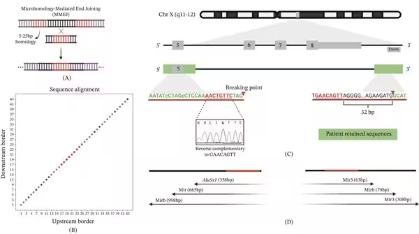
Figure 2: Microhomology at the breakpoints of the AR deletion. (A) Schematic of a microhomology‐dependent mechanism, such as microhomology‐mediated end joining (MMEJ), which utilizes short homologous sequences (5–25 bp) to mediate end joining at the break site, often resulting in deletions. (B) Dot plot alignment of upstream and downstream breakpoint borders reveals a region of microhomology (red), consistent with a microhomology‐dependent repair process. (C) Genomic context of the AR gene on chromosome X, showing a deletion spanning Exons 6–8. A microhomology of 8 bp (GAACAGTT) was identified 3 bp upstream of the 5 ′ breakpoint and 32 bp downstream (within the deleted region) on the 3 ′ side in reverse complement orientation, consistent with a microhomology‐mediated mechanism. (D) Alu and MIR elements are present near the breakpoints but distant from the junction, suggesting an indirect role in promoting genomic instability and susceptibility to microhomology‐dependent repair.
Microhomology at the breakpoints of the AR deletion. (A) Schematic of a microhomology‐dependent mechanism, such as microhomology‐mediated end joining (MMEJ), which utilizes short homologous sequences (5–25 bp) to mediate end joining at the break site, often resulting in deletions. (B) Dot plot alignment of upstream and downstream breakpoint borders reveals a region of microhomology (red), consistent with a microhomology‐dependent repair process. (C) Genomic context of the AR gene on chromosome X, showing a deletion spanning Exons 6–8. A microhomology of 8 bp (GAACAGTT) was identified 3 bp upstream of the 5 ′ breakpoint and 32 bp downstream (within the deleted region) on the 3 ′ side in reverse complement orientation, consistent with a microhomology‐mediated mechanism. (D) Alu and MIR elements are present near the breakpoints but distant from the junction, suggesting an indirect role in promoting genomic instability and susceptibility to microhomology‐dependent repair. Credit: Juliana Moreira Marques et al. / Human mutation

What These Findings Mean for Genetic Diagnostics

Standard genetic testing workflows often rely on sequencing methods that easily miss large structural deletions or duplications unless specialized algorithms are applied. The authors emphasize that integrating copy number variation screening and precise breakpoint mapping into routine clinical diagnostics can significantly improve diagnostic yields for patients with unresolved differences in sex development.

Recognizing the exact DNA repair signatures involved also sheds light on the genomic vulnerability of the X chromosome. Future clinical assessments will likely benefit from combining exome sequencing with targeted deletion screening to ensure that pathogenic structural variants are caught early in the evaluation process.

The research was published in Human mutation on January 1, 2026.

Fact Checked

This article has been fact checked for accuracy, with information verified against reputable sources. Learn more about us and our editorial process.

Last reviewed on .

Article history

  • Latest version

Reference(s)

  1. Marques, Juliana Moreira., et al. “Microhomology‐Associated Androgen Receptor Deletion and Enrichment of AR Copy‐Number Variants in Androgen Insensitivity Syndrome.” Human Mutation, vol. 2026, no. 1, November 13, 2026 Wiley, doi: 10.1155/humu/3178183. <https://doi.org/10.1155/humu/3178183>.

Cite this page:

Taylor, Elizabeth. “Hidden Chromosome Deletions Drive Severe Androgen Insensitivity Syndrome.” BioScience. BioScience ISSN 2521-5760, 16 November 2026. <https://www.bioscience.com.pk/en/subject/genetics/microhomology-associated-androgen-receptor-deletion-and-enrichment-of-ar-copy-number-variants-in-androgen-insensitivity-syndrome>. Taylor, E. (2026, November 16). “Hidden Chromosome Deletions Drive Severe Androgen Insensitivity Syndrome.” BioScience. ISSN 2521-5760. Retrieved November 16, 2026 from https://www.bioscience.com.pk/en/subject/genetics/microhomology-associated-androgen-receptor-deletion-and-enrichment-of-ar-copy-number-variants-in-androgen-insensitivity-syndrome Taylor, Elizabeth. “Hidden Chromosome Deletions Drive Severe Androgen Insensitivity Syndrome.” BioScience. ISSN 2521-5760. https://www.bioscience.com.pk/en/subject/genetics/microhomology-associated-androgen-receptor-deletion-and-enrichment-of-ar-copy-number-variants-in-androgen-insensitivity-syndrome (accessed November 16, 2026).
End of the article