Common Cellular Protein Found to Help Wake Up Dormant HIV-1
Researchers discover a metabolic and epigenetic pathway that controls viral latency, offering new clues for keeping the virus locked down.
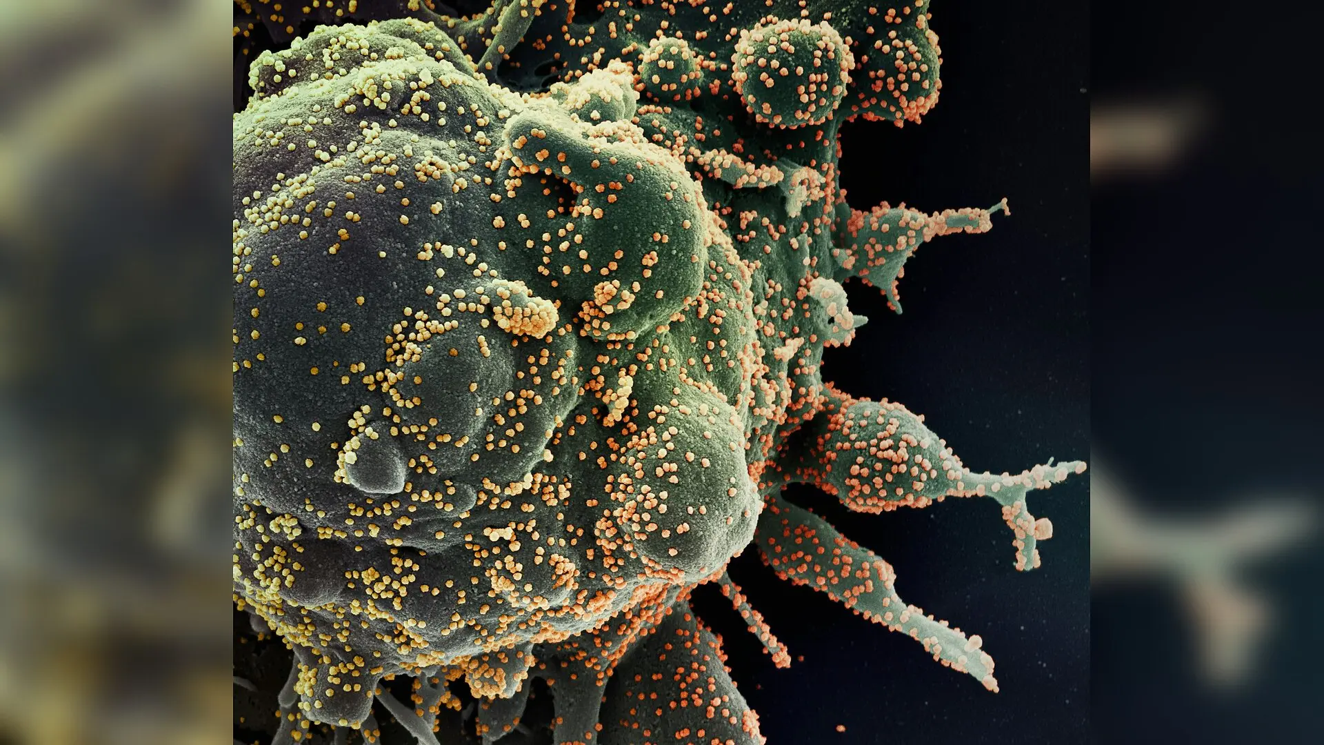
A common metabolic protein inside human cells plays a surprising role in helping human immunodeficiency virus type 1 (HIV-1) wake up from its dormant state, according to new research published in PLOS Pathogens. When scientists depleted this protein in laboratory models, viral replication and reactivation dropped significantly, pointing to a potential new avenue for keeping the virus locked down.
How Glucose and Metabolism Influence Viral Latency
While modern antiretroviral therapy successfully suppresses HIV-1 replication in patients, the virus establishes a stable, dormant reservoir inside long-lived memory T cells. This latent reservoir remains the primary barrier to a cure because stopping medication almost always triggers a rapid viral rebound. Efforts to purge this reservoir using latency-reversing agents have yielded inconsistent results, prompting researchers to explore block-and-lock strategies that reinforce viral silence without triggering widespread immune activation.
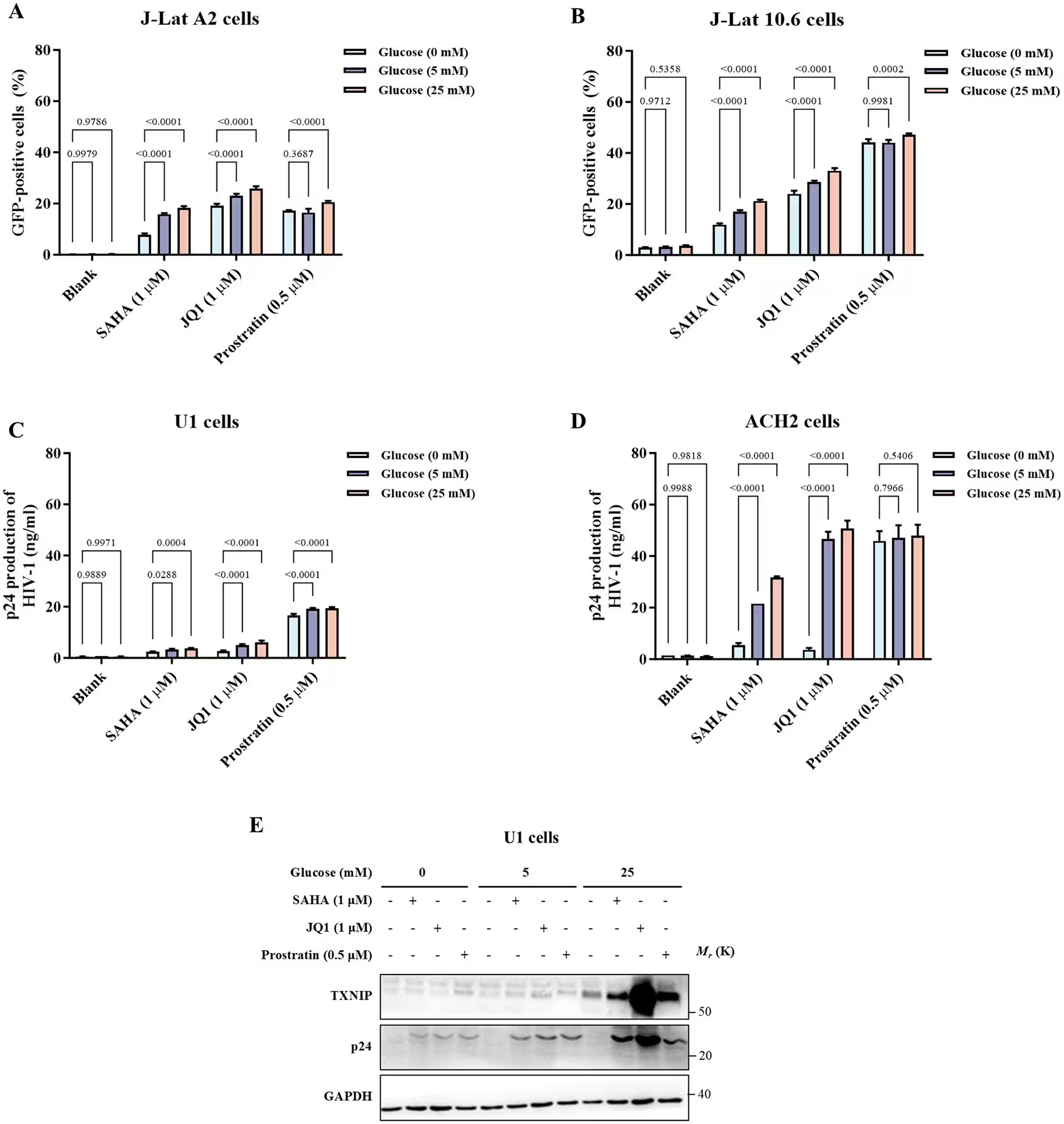
Previous studies noted that latently infected cells exhibit altered glucose handling and rely on antioxidant defenses to maintain their quiet state. Building on these clues, researchers led by a team of scientists investigated thioredoxin-interacting protein, a glucose-responsive host factor known to regulate cellular sugar uptake. Until now, the exact role of this protein in controlling viral transcription and latency remained unmapped.
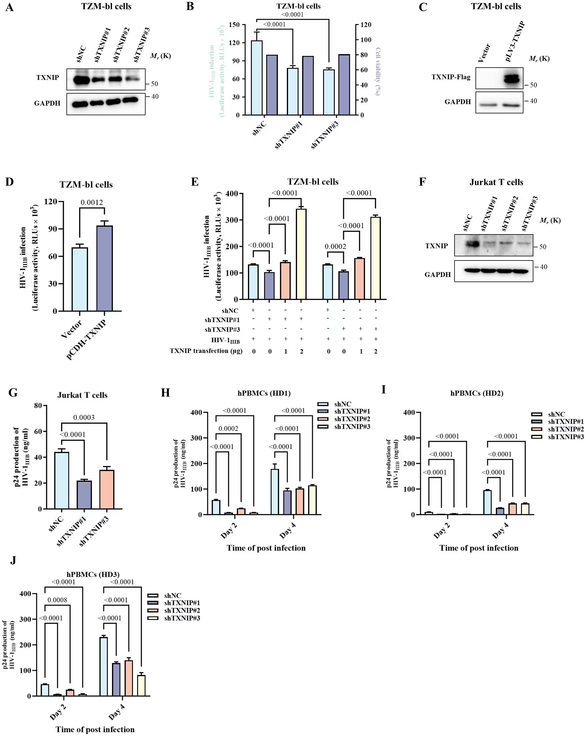
Experimental Findings in Cell Lines and Primary T Cells
The investigators tested the function of the protein across multiple experimental settings, including latently infected cell lines and primary cells isolated from healthy donors and individuals living with HIV-1 who were receiving suppressive therapy. When they silenced the gene encoding the protein using targeted ribonucleic acid interference, they observed an approximately 36 to 50 percent drop in viral particle release and lower transcriptional activity at the viral promoter region.
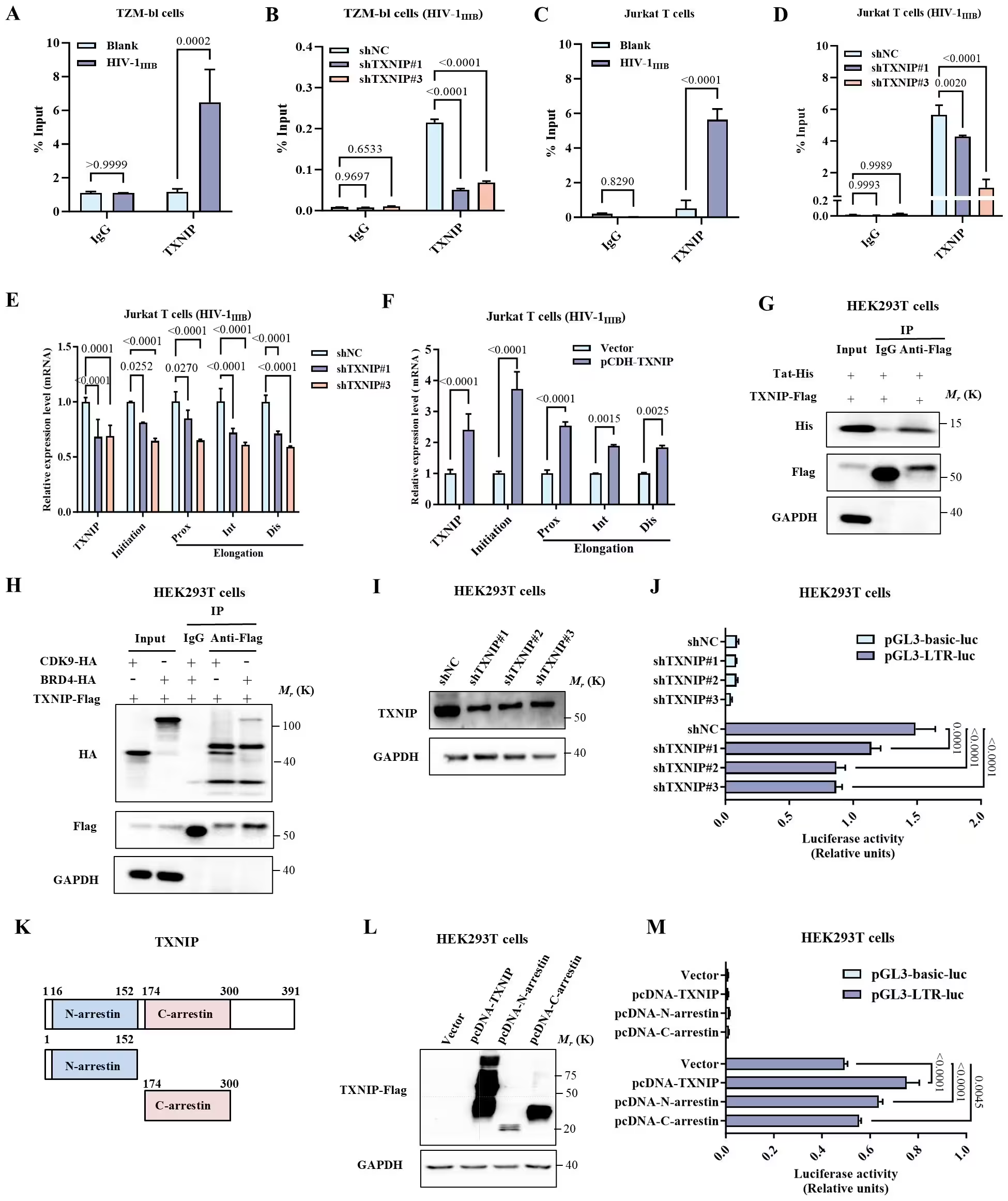
Conversely, artificially increasing the expression of the protein boosted viral transcription and enhanced the effectiveness of latency-reversing drugs. Chromatin immunoprecipitation assays further revealed that the protein associates with viral deoxyribonucleic acid sequences and interacts with cellular transcriptional machinery such as bromodomain-containing protein 4 and cyclin-dependent kinase 9, suggesting it acts as a cofactor in driving viral gene expression.
Uncovering an Epigenetic Regulatory Circuit
To understand what controls the protein, the team examined an epigenetic silencer known as enhancer of zeste homolog 2, which acts as the catalytic subunit of polycomb repressive complex 2. Elevated glucose levels decreased the abundance of this silencer, while pharmacological inhibition or genetic knockdown of the silencer increased the production of the metabolic protein and promoted viral reactivation.
Further analysis showed that this epigenetic control operates indirectly through a transcription factor designated as ETV1. The researchers found that the silencer normally represses the ETV1 gene by depositing repressive histone marks. When that repression is lifted, ETV1 binds directly to the promoter region of the metabolic protein, driving up its expression and facilitating viral transcription.
Testing a Small Molecule Probe for Viral Suppression
To evaluate whether this pathway can be targeted pharmacologically, the team tested a small-molecule compound known as SRI-37330, which binds to the metabolic protein. In infection assays across multiple viral strains and cell types, the compound suppressed viral replication with half-maximal inhibitory concentration values ranging from 0.23 to 2.80 micromolars. It also reduced virus reactivation when cells were stimulated with latency-reversing agents.
Surface plasmon resonance and cellular thermal shift assays confirmed direct target engagement between the compound and the protein. Furthermore, the suppressive effect of the compound was attenuated in cells where the protein had been genetically depleted, indicating that the protein contributes to the pharmacological activity of the drug. Cytotoxicity assays showed favorable selectivity indices, and flow cytometry analysis of peripheral blood mononuclear cells indicated no obvious induction of immune activation markers.
Study Limitations and Next Steps
The authors note that the research was conducted primarily in laboratory models and ex vivo primary cells, which cannot fully capture the complexity of viral reservoirs in living humans. Additionally, the small-molecule compound used in the experiments was originally investigated in metabolic research settings, meaning its safety and pharmacological behavior in HIV-infected individuals require thorough clinical evaluation. Future studies involving humanized animal models and broader clinical cohorts will be necessary to determine whether targeting this metabolic and epigenetic axis can translate into effective therapies for maintaining durable viral remission.
The research was published in PLOS Pathogens on November 16, 2026.
This article has been fact checked for accuracy, with information verified against reputable sources. Learn more about us and our editorial process.
Last reviewed on .
Article history
- Latest version
Reference(s)
- Wu, Ziyao., et al. “TXNIP contributes to HIV-1 transcriptional competence and inducible reactivation through an EZH2-ETV1 regulatory pathway.” PLOS Pathogens, vol. 22, no. 11, November 16, 2026, pp. e1014748 Public Library of Science (PLoS), doi: 10.1371/journal.ppat.1014748. <https://doi.org/10.1371/journal.ppat.1014748>.
Cite this page:
- Posted by Bilal Abbasi