Painless Microneedle Patches Track Disease Biomarkers Without Drawing Blood
Biotechnology

Painless Microneedle Patches Track Disease Biomarkers Without Drawing Blood

Engineered skin patches use ultrabright nanolabels to detect low-abundance immune and kidney markers directly from interstitial fluid.
By Robert Brown
Published:
Email this Article
Painless Microneedle Patches Track Disease Biomarkers Without Drawing Blood
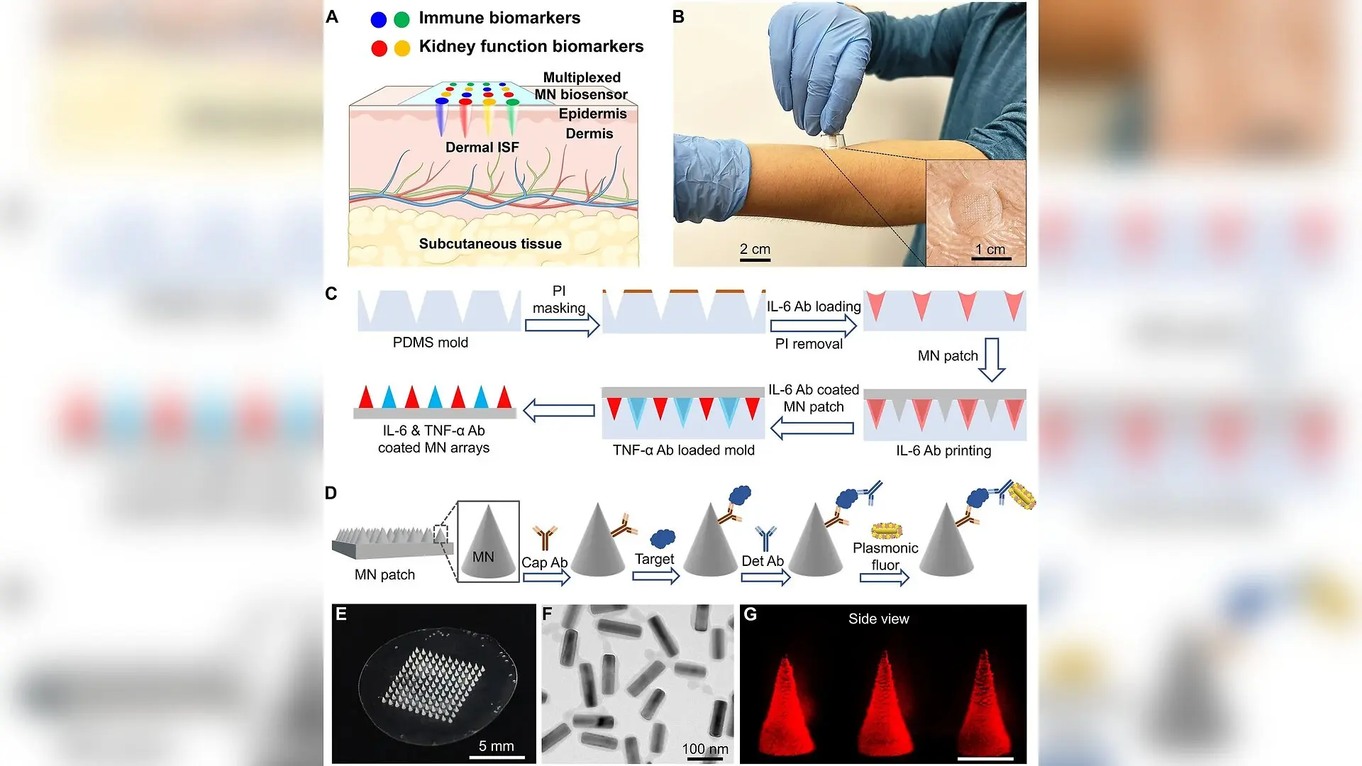
Design and fabrication of an MN patch for multiplexed biomarker detection. (A) Schematic illustration of a multiplexed MN patch enabling painless, blood-free detection, and quantification of immune and kidney function biomarkers from dermal ISF. Created in BioRender. Tian, L. (2026) https://BioRender.com/03hw88z. (B) Photograph of a human subject self-applying a multiplexed MN patch using an MN applicator. The inset image shows the patch applied to the skin covered with a medical adhesive. (C) Schematic illustration of the transfer printing of distinct antibodies on individual MNs of a single MN patch. PDMS, PI, and Ab denote polydimethylsiloxane, polyimide, and antibody, respectively. (D) Workflow of p-FLISA implemented on an MN patch for on-needle analysis of protein biomarkers. Cap Ab and Det Ab denote capture antibody and detection antibody, respectively. (E) Optical image of an MN patch consisting of an 11 × 11 MN array. (F) TEM image of PFs. (G) Side-view fluorescence microscopy Ravi Jada et al. / Science advances

Tiny needles that painlessly sample fluid just beneath the skin could soon replace blood draws for monitoring complex diseases, thanks to a novel device developed by researchers. The wearable patch successfully tracks multiple immune and kidney health markers simultaneously by combining microscopic needles with ultra-bright light-emitting labels.

Detecting multiple low-abundance protein biomarkers at the same time has long challenged biomedical engineers because tiny samples lead to cross-reactivity and binding competition. To solve this problem, the research team created a high-resolution three-dimensional transfer-pumping strategy. This method uses a soft silicone microwell array as an antibody holder to coat individual microneedles with distinct capture antibodies without cross-contamination. Each polystyrene patch comprises an 11 by 11 array of conical needles measuring 300 micrometers in base diameter and 600 micrometers in height.

To achieve exceptional analytical sensitivity down to the femtogram per milliliter range, the investigators integrated plasmonic-enhanced fluorescent nanolabels. These labels consist of gold-silver core-shell nanocuboids coated with a near-infrared fluorophore and streptavidin. The specialized nanolabels are roughly 10,000 times brighter than conventional fluorophores, allowing the biosensors to quantify very low concentrations of proteins. The work was published in Science Advances, detailing experiments across both animal models and human subjects.

How the Microneedle Patch Measures Biomarkers Without Pain

When applied to the skin using a spring-loaded applicator, the patch gently stretches the area to create a taut surface and inserts the needles into the dermal layer within a single uniform motion. The needles penetrate the epidermis and reach the upper dermis without hitting blood vessels or nerve endings, ensuring a completely blood-free and painless experience. Dermal interstitial fluid, which is the fluid surrounding tissue cells, floods the needles during a brief five-minute application window.
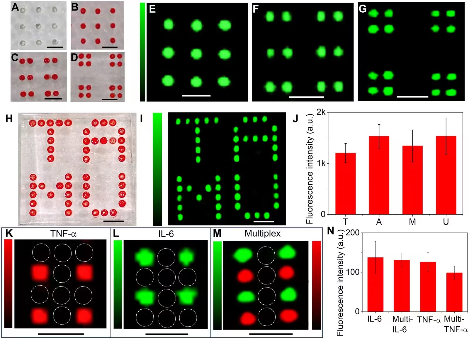
Fig. 2: Spatially controlled antibody printing on 3D MN arrays. (A to D) Optical images of PDMS antibody holder patterns and their corresponding dye-filled microwells and (E to G) fluorescence images of MN patches printed with IL-6 capture antibodies in matching patterns following p-FLISA. (H) Optical image of red dye–filled PDMS microwells in a “TAMU” pattern and (I) the corresponding fluorescence image of the MN patch printed with the same IL-6 antibody pattern. (J) Fluorescence intensities of MNs for each letter (mean ± SD, n ≥ 9). a.u., arbitrary units. Fluorescence images of MN patches with (K) alternately printed TNF-α antibodies and (L) alternately printed IL-6 antibodies following p-FLISA, respectively. White circles mark unprinted MNs. (M) Fluorescence image of a multiplexed MN patch with alternating IL-6 and TNF-α antibody patterns, with TNF-α shown in red and IL-6 shown in green. (N) Comparison of fluorescence intensities for IL-6 and TNF-α detection in singleplex and multiplexed fo
Spatially controlled antibody printing on 3D MN arrays. (A to D) Optical images of PDMS antibody holder patterns and their corresponding dye-filled microwells and (E to G) fluorescence images of MN patches printed with IL-6 capture antibodies in matching patterns following p-FLISA. (H) Optical image of red dye–filled PDMS microwells in a “TAMU” pattern and (I) the corresponding fluorescence image of the MN patch printed with the same IL-6 antibody pattern. (J) Fluorescence intensities of MNs for each letter (mean ± SD, n ≥ 9). a.u., arbitrary units. Fluorescence images of MN patches with (K) alternately printed TNF-α antibodies and (L) alternately printed IL-6 antibodies following p-FLISA, respectively. White circles mark unprinted MNs. (M) Fluorescence image of a multiplexed MN patch with alternating IL-6 and TNF-α antibody patterns, with TNF-α shown in red and IL-6 shown in green. (N) Comparison of fluorescence intensities for IL-6 and TNF-α detection in singleplex and multiplexed fo Credit: Ravi Jada et al. / Science advances

The system relies on an on-needle assay where target proteins are captured directly on the needles, followed by incubation with biotinylated detection antibodies and the ultrabright nanolabels. In laboratory evaluations, the platform quantified immune cytokines such as interleukin-6 and tumor necrosis factor-alpha alongside renal markers including neutrophil gelatinase-associated lipocalin and cystatin C. The technique yielded limits of detection in the femtogram per milliliter range for cytokines, outperforming standard enzyme-linked immunosorbent assay methods by orders of magnitude.

What Animal and Human Tests Revealed About Tracking Disease

To test how the system performs during active inflammation and organ stress, researchers administered lipopolysaccharide to mice to induce sepsis-associated acute kidney injury. Applying the microneedles before and after the injection revealed sharp spikes in inflammatory cytokines and kidney damage indicators within the interstitial fluid. Skin recovery was rapid, with tiny insertion micropores closing entirely within thirty minutes and leaving no signs of bleeding or irritation.
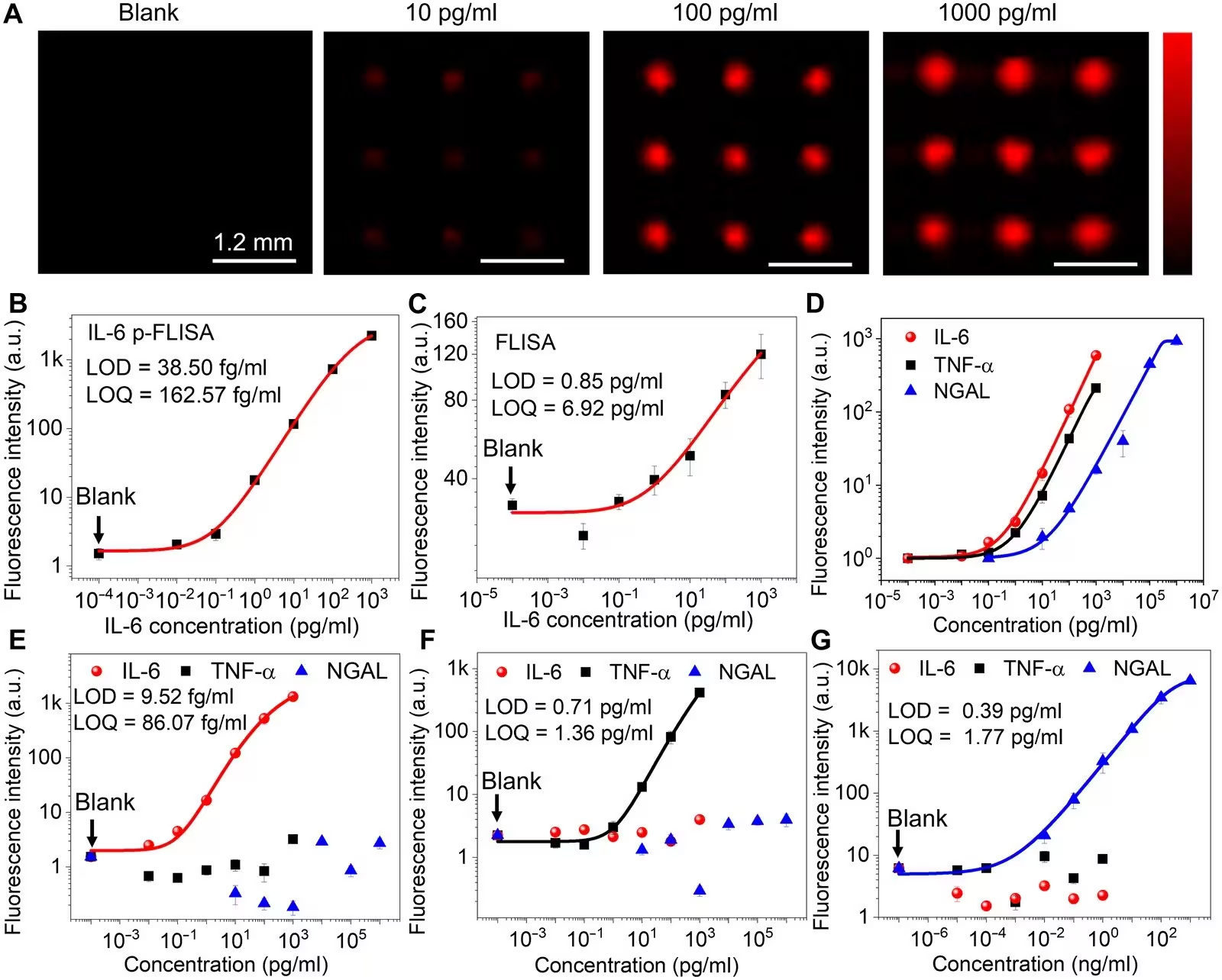
Fig. 3: Analytical performance of p-FLISA on MN patches. (A) Fluorescence images of MN patches exposed to increasing IL-6 concentrations following p-FLISA. Dose-dependent fluorescence intensities of MNs for IL-6 measured by (B) p-FLISA and (C) conventional FLISA. (D) Standard curves obtained from multiplexed MN patches for IL-6, TNF-α, and NGAL. Dose-dependent fluorescence intensities of MNs functionalized with (E) IL-6, (F) TNF-α, and (G) NGAL capture antibodies following exposure to IL-6, TNF-α, and NGAL, respectively, and subsequent p-FLISA. Data in (B) to (G) are presented as mean ± SD (n = 4).
Analytical performance of p-FLISA on MN patches. (A) Fluorescence images of MN patches exposed to increasing IL-6 concentrations following p-FLISA. Dose-dependent fluorescence intensities of MNs for IL-6 measured by (B) p-FLISA and (C) conventional FLISA. (D) Standard curves obtained from multiplexed MN patches for IL-6, TNF-α, and NGAL. Dose-dependent fluorescence intensities of MNs functionalized with (E) IL-6, (F) TNF-α, and (G) NGAL capture antibodies following exposure to IL-6, TNF-α, and NGAL, respectively, and subsequent p-FLISA. Data in (B) to (G) are presented as mean ± SD (n = 4). Credit: Ravi Jada et al. / Science advances

Human trials involved six healthy participants aged 20 to 70 who self-applied the patches to their forearms using the mechanical applicator. All participants reported zero pain or discomfort, and their skin recovered fully within half an hour. Biomarker concentrations measured in the skin fluid showed strong statistical correlations with matching blood plasma levels collected at the same time, confirming that the microneedle approach accurately reflects systemic health conditions.

Overcoming Manufacturing Hurdles for Future Clinical Use

Despite these promising results, several hurdles remain before the patches can become widely available clinical tools. The current manual transfer-printing process requires precise alignment and skilled handling, meaning that automated manufacturing will be necessary to scale up production and reduce batch-to-batch variation. Additionally, researchers must develop ambient preservation strategies to extend the shelf life of the antibody-coated patches so they can be stored without relying on continuous cold-chain refrigeration.

Future development will focus on streamlining automated fabrication and testing the sensors in larger patient cohorts experiencing acute clinical conditions. If successful, the technology could pave the way for frequent, wearable disease monitoring and personalized medicine in point-of-care settings.

The research was published in Science advances on November 18, 2026.

Fact Checked

This article has been fact checked for accuracy, with information verified against reputable sources. Learn more about us and our editorial process.

Last reviewed on .

Article history

  • Latest version

Reference(s)

  1. Jada, Ravi., et al. “Spatially multiplexed microneedle biosensors for low-abundance protein biomarker monitoring in dermal interstitial fluid.” Science Advances, vol. 12, no. 47, November 20, 2026 American Association for the Advancement of Science (AAAS), doi: 10.1126/sciadv.aeh9246. <https://doi.org/10.1126/sciadv.aeh9246>.

Cite this page:

Brown, Robert. “Painless Microneedle Patches Track Disease Biomarkers Without Drawing Blood.” BioScience. BioScience ISSN 2521-5760, 21 November 2026. <https://www.bioscience.com.pk/en/subject/biotechnology/spatially-multiplexed-microneedle-biosensors-for-low-abundance-protein-biomarker-monitoring-in-dermal-interstitial-fluid>. Brown, R. (2026, November 21). “Painless Microneedle Patches Track Disease Biomarkers Without Drawing Blood.” BioScience. ISSN 2521-5760. Retrieved November 21, 2026 from https://www.bioscience.com.pk/en/subject/biotechnology/spatially-multiplexed-microneedle-biosensors-for-low-abundance-protein-biomarker-monitoring-in-dermal-interstitial-fluid Brown, Robert. “Painless Microneedle Patches Track Disease Biomarkers Without Drawing Blood.” BioScience. ISSN 2521-5760. https://www.bioscience.com.pk/en/subject/biotechnology/spatially-multiplexed-microneedle-biosensors-for-low-abundance-protein-biomarker-monitoring-in-dermal-interstitial-fluid (accessed November 21, 2026).
End of the article