Thin Gum Tissue Triggers Greater Bone Loss Around Dental Implants
A one-year study reveals how soft tissue thickness directly influences crestal bone stability following immediate implant placement in the upper jaw.
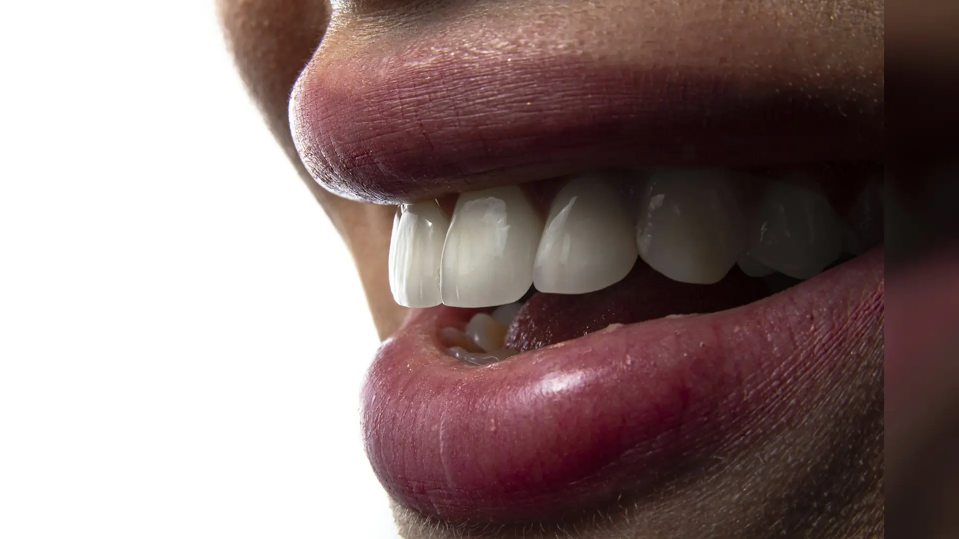
Patients with thin gums face a much higher risk of bone loss around dental implants placed immediately after tooth extraction in the front of the upper jaw, according to a retrospective cohort study. The new research demonstrates that soft tissue thickness acts as a powerful predictor of how much jawbone will remain stable during the first year of functional chewing.
Dental implants placed in the anterior maxilla, which is the front region of the upper jaw, must satisfy demanding cosmetic and structural requirements. Even minor bone resorption in this visible zone can compromise the appearance of the gums and teeth. Researchers have long debated whether the thickness of the gum tissue surrounding an implant dictates how much underlying bone will dissolve during the healing and loading phases.
To investigate this relationship, scientists analyzed clinical records from 50 patients who received single-tooth implants immediately following extraction. The study was published in the journal PLOS One by researchers Mahdi Kadkhodazadeh, Mostafa Alam, Mohsen Golkar, and Tara Naghshbandi. All participants had healthy systems with intact facial bone walls and received complete follow-up examinations one year after functional loading.
How Gum Measurements Reveal Implant Risks
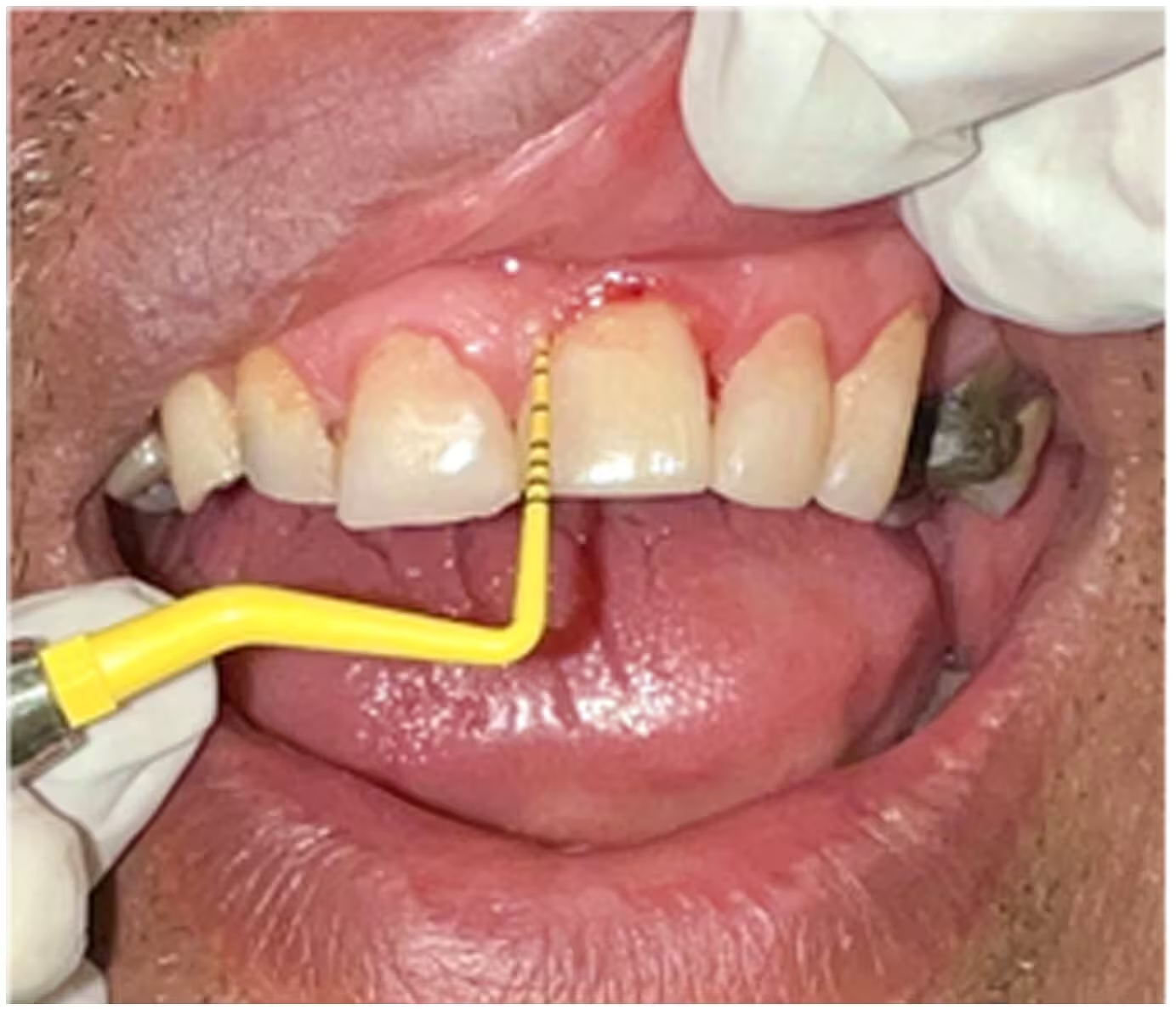
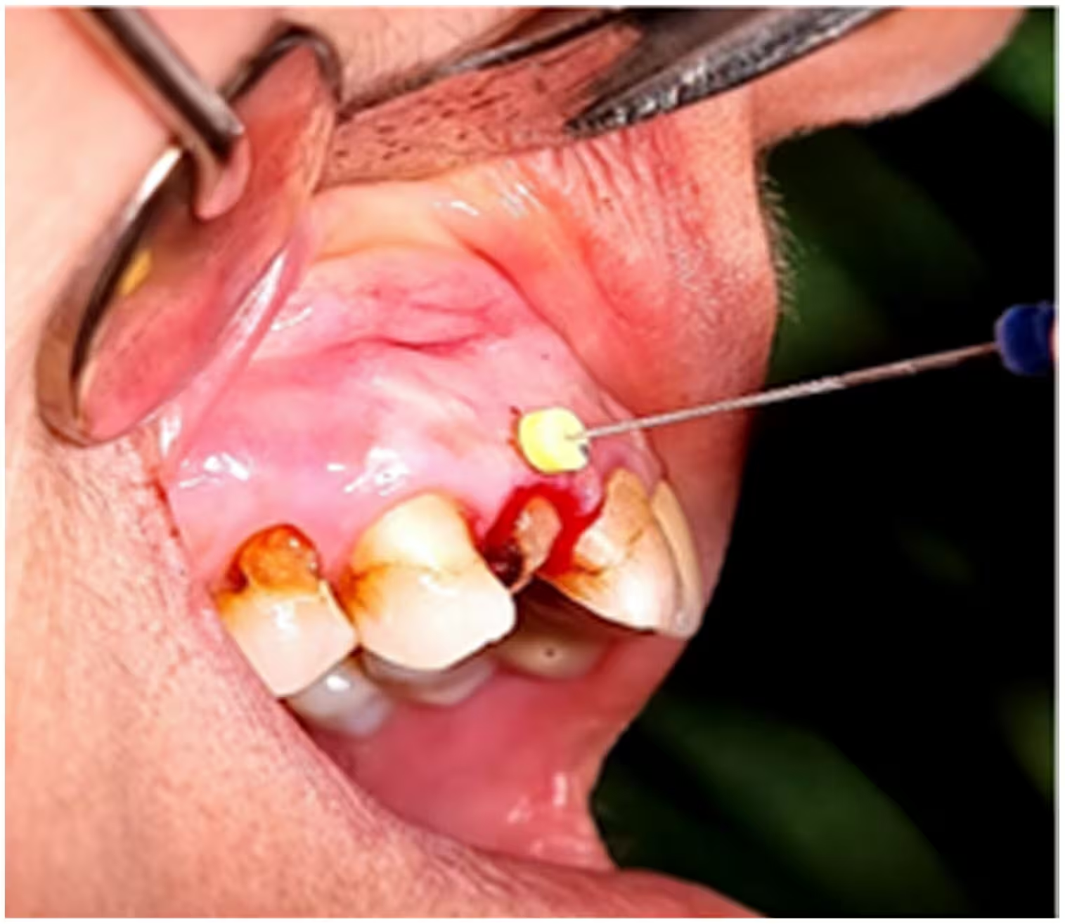
During the surgical procedures, clinicians measured the mid-buccal soft tissue thickness, which is the depth of the gum tissue on the cheek side of the implant site, using an endodontic file inserted three millimeters below the gum margin. Based on these measurements, the team divided the 50 implant sites into two distinct groups. Twenty-two sites featured a thin phenotype with soft tissue thickness measuring one millimeter or less, while 28 sites possessed a thick phenotype with soft tissue thickness exceeding one millimeter.
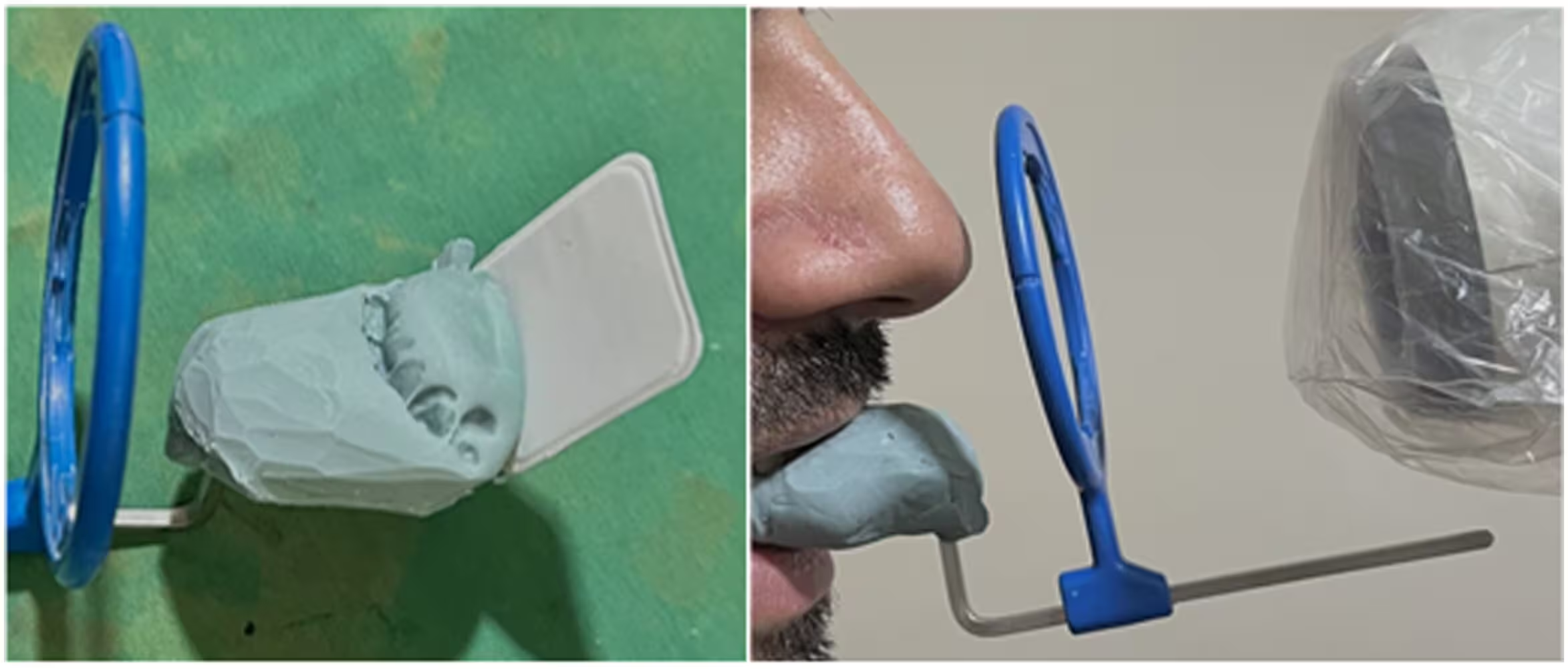
The investigators also tracked various clinical markers, including probing depth, bleeding on probing, plaque index, and keratinized tissue width. Radiographic evaluations using standardized periapical digital radiographs with custom silicone positioning indexes allowed precise measurement of marginal bone loss, defined as the vertical distance between the bone crest and the implant shoulder.
Thin Gums Trigger Greater Bone Loss
After one year of functional loading, the results revealed a stark difference in bone stability between the two phenotypes. Implants placed in thin soft tissue sites suffered an average marginal bone loss of 1.34 millimeters. In contrast, implants surrounded by thick soft tissue experienced an average bone loss of just 1.00 millimeter, representing a statistically significant difference.
Further statistical evaluation using Spearman rank correlation confirmed a strong negative relationship between soft tissue thickness and marginal bone loss, with a correlation coefficient of negative 0.749. Other clinical parameters, such as plaque accumulation, bleeding percentages, and keratinized tissue width, showed no significant differences between the thin and thick groups. This indicates that gum thickness alone served as the dominant local factor influencing crestal bone stability.
Why Tissue Thickness Protects Jawbone
The biological explanation for these findings centers on the concept of supracrestal tissue attachment, which is the natural biological seal formed by epithelial and connective tissues around an implant. When the surrounding mucosa is thin, the vertical space available for this protective seal is insufficient. To re-establish the necessary biological width, the body initiates adaptive bone remodeling, causing the crestal bone to resorb downward.
Furthermore, thicker gum tissue provides better vascular density and a more robust collagen matrix, which improves nutrient exchange and mechanical buffering. Thicker tissue is better equipped to dissipate compressive forces and reduce strain accumulation at the bone-implant interface. In contrast, thin mucosal architecture leaves the underlying bone vulnerable to inflammation and resorption.
Study Limitations and Clinical Implications
The authors noted several important limitations in their study. The retrospective design restricts the findings to associations rather than direct causation. The evaluation relied on two-dimensional periapical radiographs rather than three-dimensional cone-beam computed tomography, which could provide more detailed imaging of facial bone plates. Additionally, the study did not stratify implants by internal connection design, leaving open the possibility that platform-switching features may have influenced bone outcomes.
Despite these constraints, the findings suggest that clinicians should evaluate soft tissue phenotypes prior to immediate implant placement in the esthetic zone. Identifying patients with thin gums allows dental professionals to incorporate preventive strategies, such as tissue augmentation grafts or subcrestal implant positioning, to protect long-term bone levels and preserve a natural smile.
The research was published in PLOS One on November 20, 2026.
This article has been fact checked for accuracy, with information verified against reputable sources. Learn more about us and our editorial process.
Last reviewed on .
Article history
- Latest version
Reference(s)
- Kadkhodazadeh, Mahdi., et al. “Influence of peri-implant soft tissue thickness on marginal bone loss following immediate implant placement in the anterior maxilla: A retrospective comparative study.” PLOS One, vol. 21, no. 11, November 20, 2026, pp. e0360723 Public Library of Science (PLoS), doi: 10.1371/journal.pone.0360723. <https://doi.org/10.1371/journal.pone.0360723>.
Cite this page:
- Posted by Rohan Kumar