Brain Cells Use Mobile Vesicles to Deliver Protein Factories on Demand
Biotechnology

Brain Cells Use Mobile Vesicles to Deliver Protein Factories on Demand

Advanced imaging reveals that endoplasmic reticulum-derived vesicles transport ribosomes to distant neuronal branches to support synaptic plasticity.
By Rohan Kumar
Published:
Email this Article
Brain Cells Use Mobile Vesicles to Deliver Protein Factories on Demand
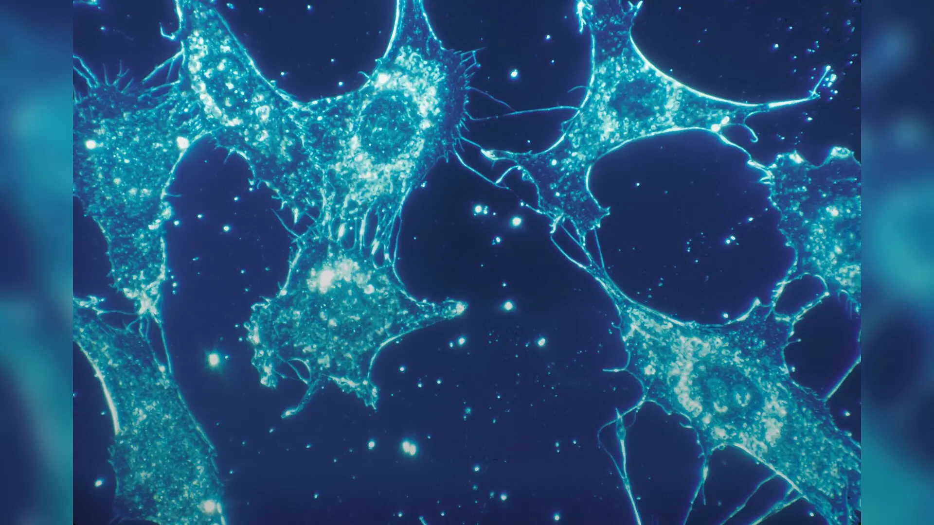
Photo by National Cancer Institute on Unsplash

Brain cells rely on tiny transport vehicles to deliver protein-building machinery directly to distant branches, solving a long-standing biological mystery. Scientists have identified specialized structures derived from the cellular recycling and folding network that carry ribosomes to faraway neuronal projections.

Neurons are exceptionally polarized cells with branching extensions called axons and dendrites that can stretch over long distances away from the main cell body. To maintain synaptic plasticity and respond swiftly to environmental cues, these remote regions carry out local protein synthesis rather than waiting for proteins to travel all the way from the center of the cell. However, researchers have long debated how ribosomes, the cellular factories that translate messenger RNA into proteins, manage to populate these distant outposts.

To investigate this process, a multi-institution research team combined advanced imaging techniques, including cryo-electron tomography and live-cell microscopy, to examine rat, mouse, and postmortem human brain tissue. The work was published in Science Advances under the title Ribosome-associated vesicles promote activity-dependent local translation in neurons. The team discovered that the endoplasmic reticulum, an interconnected membranous network typically found near the nucleus, buds off distinct vesicles that become decorated with 80S ribosomes.

How Vesicles Act as Mobile Translation Platforms

The researchers tracked these structures, termed ribosome-associated vesicles or RAVs, and found they measure approximately 400 nanometers in diameter. Using high-resolution electron imaging, the authors confirmed that these vesicles are densely coated with mammalian ribosomes arranged in orderly patterns resembling polysomes. Live-cell tracking revealed that these vesicles exhibit dynamic behaviors, with some moving rapidly along microtubule tracks while others remain stationary.
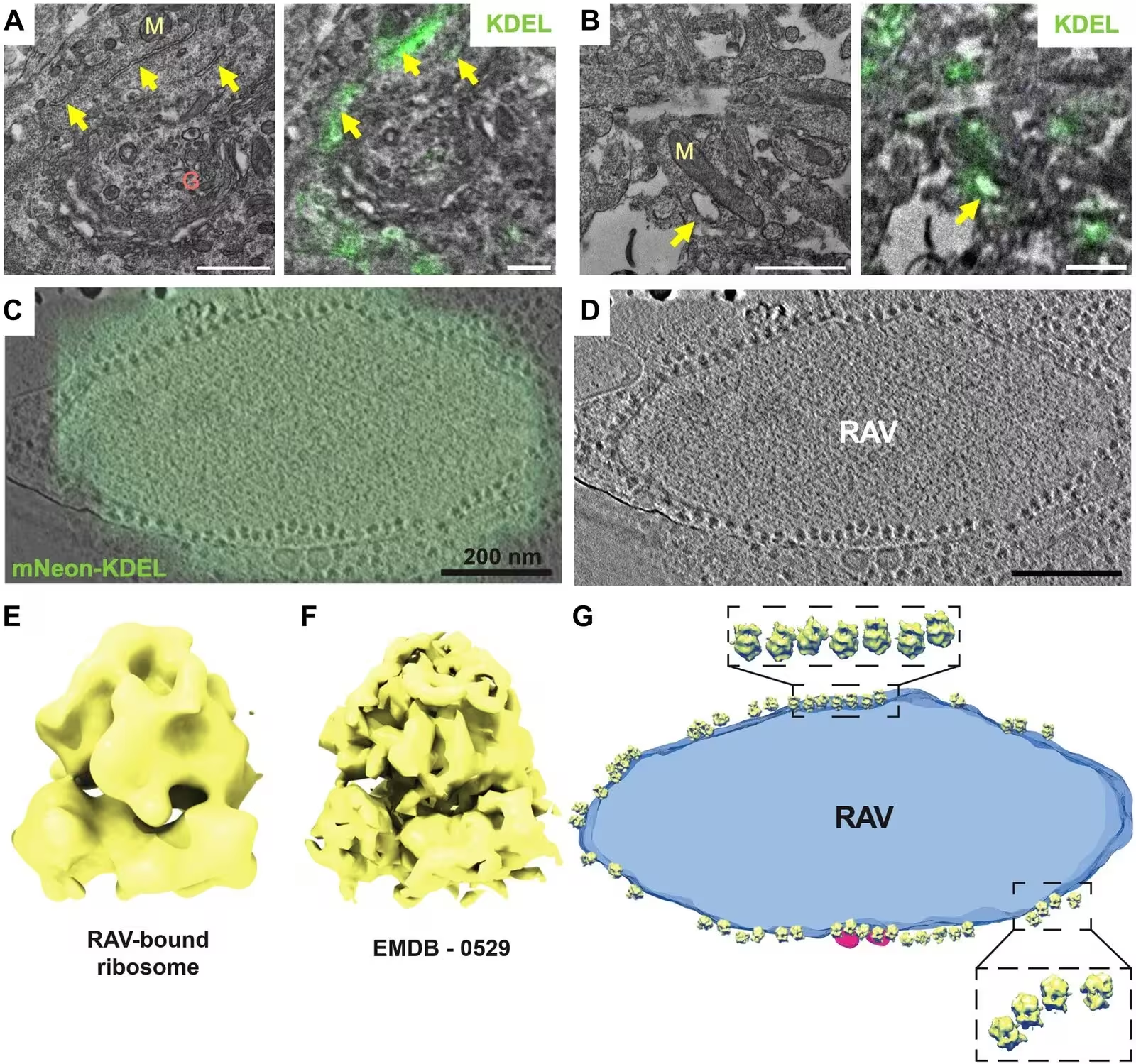
Fig. 1: Identification of RAVs by correlative light and electron microscopy (CLEM) of KDEL-labeled structures. (A and B) CLEM imaging of endoplasmic reticulum (ER) distribution in primary rat cortical neurons using an anti-KDEL antibody. (A) A representative high-magnification micrograph (left panel) demonstrated extensive ER network in the cell soma. Correlative image (right panel) showing co-localization of anti-KDEL signal (in green) with ER (arrow). The Golgi apparatus (G) and mitochondria (M) are also visible. Scale bar, 1 μm. (B) Higher magnification of regions showing a KDEL-positive vesicle. Right panel: correlative images showing the same vesicular structures co-localizing with anti-KDEL antibody. Scale bar, 500 nm. (C and D) Cryo-CLEM shows ER marker mNeon-KDEL localized to discrete puncta in the periphery of primary hippocampal neurons. A cryo-tomographic slice with (C) and without (D) overlay of an epifluorescence image with mNeon-KDEL fluorescence (in green). Scale bars, 200 nm. (
Identification of RAVs by correlative light and electron microscopy (CLEM) of KDEL-labeled structures. (A and B) CLEM imaging of endoplasmic reticulum (ER) distribution in primary rat cortical neurons using an anti-KDEL antibody. (A) A representative high-magnification micrograph (left panel) demonstrated extensive ER network in the cell soma. Correlative image (right panel) showing co-localization of anti-KDEL signal (in green) with ER (arrow). The Golgi apparatus (G) and mitochondria (M) are also visible. Scale bar, 1 μm. (B) Higher magnification of regions showing a KDEL-positive vesicle. Right panel: correlative images showing the same vesicular structures co-localizing with anti-KDEL antibody. Scale bar, 500 nm. (C and D) Cryo-CLEM shows ER marker mNeon-KDEL localized to discrete puncta in the periphery of primary hippocampal neurons. A cryo-tomographic slice with (C) and without (D) overlay of an epifluorescence image with mNeon-KDEL fluorescence (in green). Scale bars, 200 nm. ( Credit: Eva Martin-Solana et al. / Science advances

When the researchers simulated neuronal activity using a chemical protocol to induce long-term potentiation, a cellular model for learning and memory, the dynamics shifted dramatically. In dendrites, the density of these vesicles increased by roughly 60 percent, primarily driven by moving vesicles coming to a halt at specific sites. Furthermore, chemical stimulation increased the proportion of vesicles carrying ribosomes, indicating that neurons can recruit translational machinery on demand.

What Happens When Neurons Receive Stimulation

Using single-molecule imaging to monitor translation sites in real time, the team observed that stationary vesicles frequently co-localized with active translation sites in dendrites. Regions where these vesicles associated with translation sites exhibited roughly 30 percent greater signal amplitude compared to periods without vesicle association. This points to a direct role for these structures in boosting local protein output during heightened neuronal activity.
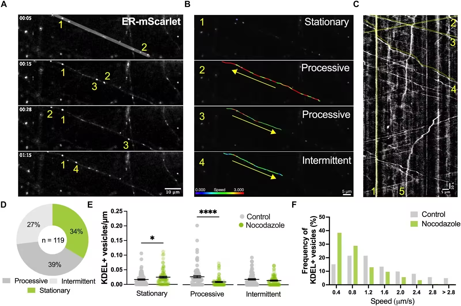
Fig. 2: KDEL-positive vesicle dynamics in neurites. (A) Representative time-lapse images demonstrate the dynamics of ER-mScarlet-labeled vesicles (numbered 1–4) over time along a neurite of a primary cortical neuron (DIV11–12). Scale bar, 10 μm. (B) Frame-by-frame velocity measurements of KDEL-positive vesicles in (A). Each of the 4 highlighted vesicles (1-4) demonstrated different dynamics as indicated by the color-coded velocity range: stationary (1), fast (2), moderate (3), and slow (4) velocities, respectively. Arrows specify the overall direction of movement along the neurite. Scale bar, 5 μm. (C) Kymograph of the segmented neurite from (A). Highlighted lines (in yellow) indicate movements of the KDEL-positive vesicles (1-4) featured in (A) and (B) (1-4) across a 5-minute timespan. (5) indicates a KDEL-positive vesicle with intermittent movement prior to stopping in place. Scale bar, 5 μm, 10-second imaging. (D) Relative proportion of moving (processive and intermittent) versus stationary
KDEL-positive vesicle dynamics in neurites. (A) Representative time-lapse images demonstrate the dynamics of ER-mScarlet-labeled vesicles (numbered 1–4) over time along a neurite of a primary cortical neuron (DIV11–12). Scale bar, 10 μm. (B) Frame-by-frame velocity measurements of KDEL-positive vesicles in (A). Each of the 4 highlighted vesicles (1-4) demonstrated different dynamics as indicated by the color-coded velocity range: stationary (1), fast (2), moderate (3), and slow (4) velocities, respectively. Arrows specify the overall direction of movement along the neurite. Scale bar, 5 μm. (C) Kymograph of the segmented neurite from (A). Highlighted lines (in yellow) indicate movements of the KDEL-positive vesicles (1-4) featured in (A) and (B) (1-4) across a 5-minute timespan. (5) indicates a KDEL-positive vesicle with intermittent movement prior to stopping in place. Scale bar, 5 μm, 10-second imaging. (D) Relative proportion of moving (processive and intermittent) versus stationary Credit: Eva Martin-Solana et al. / Science advances

The study also revealed that these vesicles interact selectively with specific messenger RNA transcripts. While association with housekeeping or structural transcripts remained stable, chemical stimulation significantly increased the recruitment of Nr2a transcripts, which encode a subunit of the NMDA glutamate receptor. This selective engagement suggests that neurons can fine-tune which proteins are manufactured locally based on specific signaling pathways.

Current Limitations and Next Steps

Despite these clear findings, the researchers noted several limitations to their approach. Because no single marker uniquely labels these specific vesicles, the team relied on the endoplasmic reticulum retention signal KDEL, which can also label other compartments like the Golgi apparatus. Additionally, heavy metal staining required for traditional volume electron microscopy lacks the resolution to definitively identify individual ribosomes, necessitating complementary cryogenic techniques.

Future investigations will need to explore the exact molecular mechanisms controlling how these vesicles form and whether ribosome-free vesicles act as a dynamic reserve pool. Uncovering how these platforms target specific mRNAs and coordinate with other cellular transport systems will further clarify the complex mechanics of neuronal communication and memory formation.

The research was published in Science advances on November 18, 2026.

Fact Checked

This article has been fact checked for accuracy, with information verified against reputable sources. Learn more about us and our editorial process.

Last reviewed on .

Article history

  • Latest version

Reference(s)

  1. Martin-Solana, Eva., et al. “Ribosome-associated vesicles promote activity-dependent local translation in neurons.” Science Advances, vol. 12, no. 47, November 20, 2026 American Association for the Advancement of Science (AAAS), doi: 10.1126/sciadv.aee0620. <https://doi.org/10.1126/sciadv.aee0620>.

Cite this page:

Kumar, Rohan. “Brain Cells Use Mobile Vesicles to Deliver Protein Factories on Demand.” BioScience. BioScience ISSN 2521-5760, 21 November 2026. <https://www.bioscience.com.pk/en/subject/biotechnology/ribosome-associated-vesicles-promote-activity-dependent-local-translation-in-neurons>. Kumar, R. (2026, November 21). “Brain Cells Use Mobile Vesicles to Deliver Protein Factories on Demand.” BioScience. ISSN 2521-5760. Retrieved November 21, 2026 from https://www.bioscience.com.pk/en/subject/biotechnology/ribosome-associated-vesicles-promote-activity-dependent-local-translation-in-neurons Kumar, Rohan. “Brain Cells Use Mobile Vesicles to Deliver Protein Factories on Demand.” BioScience. ISSN 2521-5760. https://www.bioscience.com.pk/en/subject/biotechnology/ribosome-associated-vesicles-promote-activity-dependent-local-translation-in-neurons (accessed November 21, 2026).
End of the article