Why Nails Grow and Do Not Feel Pain: The Science Behind Nail Formation and Pain-Free Trimming
Human nails are rigid keratin structures produced in the nail matrix through continuous cell division. Once formed, the nail plate becomes non-living and nerve-free, explaining painless cutting and revealing the complex biology that governs nail growth.
The nails are specialized keratinized structures derived from the epidermis and represent a unique modification of skin designed primarily for protection and fine motor function. Although nails are often regarded as simple, inert appendages, their formation, composition, and growth involve highly coordinated biochemical, cellular, and physiological processes. A detailed understanding of nail biochemistry and growth mechanisms is important not only for basic biology but also for clinical interpretation, as many systemic and local diseases manifest early changes in the nails. Furthermore, the absence of pain during nail cutting highlights fundamental principles of tissue differentiation and sensory biology.
Structural Organization of the Nail Unit
The nail unit is a complex anatomical structure composed of several distinct but functionally integrated components. These include the nail plate, nail matrix, nail bed, nail folds, cuticle or eponychium, and hyponychium. Among these, the nail plate is the visible hard structure, whereas the nail matrix is the principal site of nail production.
The nail plate rests on the nail bed and extends distally beyond the fingertip to form the free edge. Proximally, it is partially covered by the proximal nail fold. The pale crescent-shaped lunula visible in some individuals represents the distal portion of the nail matrix, where keratinization is incomplete and light reflection differs from the fully keratinized nail plate. Each component of the nail unit contributes to normal nail growth, attachment, and protection.
Biochemical Composition of Nails
Keratin as the Primary Structural Protein
The nail plate is composed predominantly of hard keratin, a fibrous structural protein belonging to the family of intermediate filament proteins. Keratin molecules are synthesized by specialized epithelial cells called keratinocytes and are characterized by a high content of sulfur-rich amino acids, particularly cysteine. The abundance of cysteine allows the formation of extensive disulfide bonds, which are covalent linkages between sulfur atoms of adjacent cysteine residues.
These disulfide bonds are critical determinants of nail hardness, rigidity, and resistance to mechanical stress. Compared with soft keratin found in the epidermis, nail keratin contains a significantly higher density of these cross-links, explaining its greater strength and reduced flexibility. Alterations in sulfur metabolism or cysteine availability can impair keratin structure and lead to brittle or fragile nails.
Keratin Filament Organization
At the molecular level, keratin proteins assemble into heterodimers, which then aggregate to form protofilaments, protofibrils, and ultimately mature intermediate filaments. These filaments are embedded in a dense matrix of keratin-associated proteins that further enhance structural stability. The highly ordered packing of keratin filaments contributes to the lamellar architecture of the nail plate, which is arranged in tightly compacted layers.
This layered organization explains why nails can split longitudinally or horizontally under pathological or environmental stress. It also accounts for the slow diffusion of substances through the nail plate, a factor of clinical relevance in topical drug delivery.
Water, Lipids, and Mineral Content
Although keratin constitutes the majority of nail mass, nails also contain approximately 10 to 30 percent water. The water content influences nail flexibility and tensile strength. Dehydration leads to brittle nails, whereas excessive hydration softens the nail plate. Lipids, including cholesterol, phospholipids, and free fatty acids, are present in small amounts and play a role in maintaining nail barrier function.
Trace elements such as zinc, iron, copper, selenium, and magnesium are detectable in nails. These elements are incorporated during nail formation and reflect systemic nutritional and metabolic status. Calcium is often believed to strengthen nails, but it is not a major structural component of nail keratin. Instead, minerals act as cofactors for enzymes involved in keratin synthesis and cellular metabolism within the nail matrix.
Cellular Biology of Nail Formation
The Nail Matrix as a Proliferative Zone
The nail matrix is a highly specialized germinative epithelium responsible for producing the nail plate. It contains rapidly dividing keratinocytes with high metabolic activity. These cells are nourished by a rich capillary network and are under tight regulatory control by growth factors, hormones, and local signaling pathways.
Keratinocytes in the matrix undergo continuous mitosis. As new cells are produced, older cells are pushed distally and upward. During this migration, keratinocytes begin to synthesize large quantities of keratin proteins and keratin-associated proteins.
Keratinization Process
Keratinization in the nail matrix differs from epidermal keratinization. In the skin, keratinocytes form a granular layer containing keratohyalin granules, whereas in the nail matrix, this granular layer is absent or minimal. Instead, keratinization is more compact and direct, leading to the formation of dense, hard keratin.
As matrix keratinocytes differentiate, they progressively lose their nuclei, mitochondria, ribosomes, and other organelles. Cellular metabolism ceases, and the cells become flattened, anucleate, and tightly packed. The end result is the formation of the nail plate, which is composed entirely of dead, keratinized cells.
Mechanism of Nail Growth
Direction and Rate of Growth
Nail growth is a continuous process driven by sustained cell division in the nail matrix. The nail plate moves distally over the nail bed as newly formed keratinized cells accumulate beneath it. The nail bed itself contributes minimally to nail plate thickness but plays an important role in adhesion and support.
Fingernails grow at an average rate of approximately 0.1 millimeters per day, equivalent to about 3 millimeters per month. Toenails grow more slowly, often at half this rate. Complete replacement of a fingernail takes about 4 to 6 months, whereas toenails may require 12 to 18 months for full regrowth.
Factors Influencing Nail Growth
Nail growth rate varies considerably among individuals and is influenced by multiple factors. Age is an important determinant, with faster growth observed in children and young adults and slower growth in the elderly. Nutritional status, particularly protein intake and micronutrient availability, plays a critical role in supporting matrix cell proliferation.
Hormonal factors also influence nail growth. Thyroid hormones stimulate keratinocyte metabolism, and hyperthyroidism is associated with accelerated nail growth, whereas hypothyroidism results in slow-growing, brittle nails. Pregnancy is often associated with increased nail growth due to hormonal and circulatory changes.
Local factors such as trauma, inflammation, and blood supply can alter growth. Increased blood flow to the fingers, as seen in warmer climates or during physical activity, is associated with faster nail growth. Conversely, systemic illness, fever, or severe stress can temporarily arrest matrix activity, producing transverse grooves known as Beau’s lines.
Sensory Biology and the Absence of Pain During Nail Cutting
Lack of Innervation in the Nail Plate
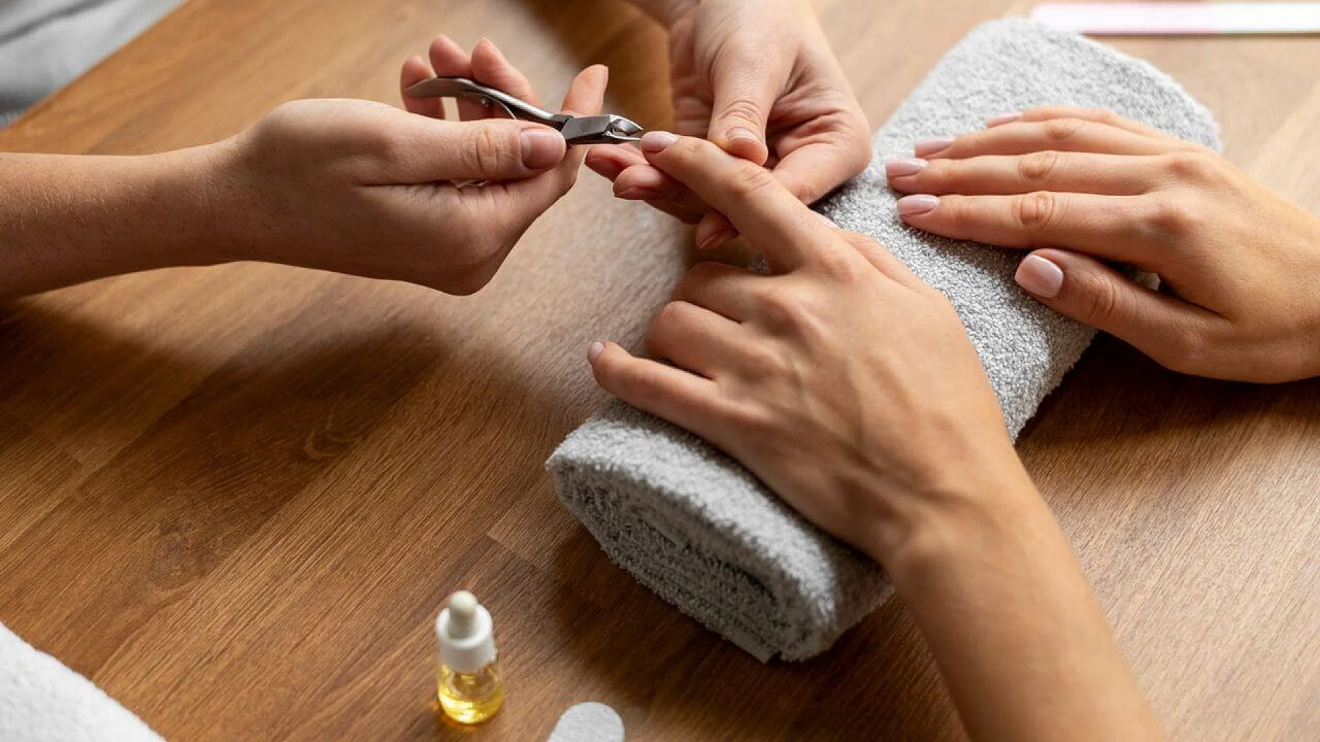
Pain perception requires living tissue equipped with sensory nerve endings capable of transducing mechanical, thermal, or chemical stimuli into neural signals. The nail plate, being composed entirely of dead, fully keratinized cells, lacks blood vessels, lymphatics, and nerves. As a result, cutting, trimming, or filing the nail plate does not activate nociceptors and is therefore painless.
This property of nails is analogous to hair shafts, which are also keratinized and non-innervated once they emerge from the follicle. Sensory perception is confined to the living tissues from which these structures originate.
Pain Sensitivity of Surrounding Structures
In contrast to the nail plate, the nail bed, nail matrix, and periungual skin are richly innervated and highly sensitive. These tissues contain free nerve endings, mechanoreceptors, and nociceptors that respond to injury or inflammation. When nail cutting extends beyond the free edge into the nail bed or hyponychium, pain and bleeding occur due to damage to these living structures.
The nail matrix is particularly sensitive, and injury to this region can result in severe pain and permanent nail deformities. This explains why deep nail trauma is both painful and clinically significant, whereas superficial trimming is not.
Clinical Correlations and Diagnostic Significance
Nails as Indicators of Systemic Health
Because nails grow slowly and incorporate biochemical information during formation, they serve as valuable indicators of systemic health. Changes in nail color, shape, thickness, or surface texture often reflect underlying nutritional deficiencies, metabolic disorders, or systemic diseases.
For example, iron deficiency may cause spoon-shaped nails, protein deficiency can lead to brittle nails, and chronic liver disease may produce white or pale nails. The presence of transverse lines, discoloration, or abnormal growth patterns provides insight into the timing and severity of systemic insults.
Drug and Toxicological Implications
Nails can accumulate drugs, heavy metals, and environmental toxins during growth. This property is utilized in forensic and toxicological analysis to assess chronic exposure. Unlike blood or urine, nails provide a long-term record of exposure, as incorporated substances remain trapped within the keratin matrix until the nail grows out.
Nutritional and Metabolic Considerations
Adequate nail formation requires sufficient intake of proteins, sulfur-containing amino acids, vitamins, and trace elements. Biotin plays a role in keratin infrastructure and cellular metabolism, and deficiency is associated with brittle nails. Zinc is essential for DNA synthesis and cell division in the nail matrix, while iron supports oxygen delivery to rapidly dividing cells.
However, excessive supplementation does not necessarily improve nail strength in individuals with normal nutritional status. Nail health is best maintained through balanced nutrition and proper care rather than pharmacological intervention.
Conclusion
The nails are complex biological structures formed through a highly regulated process of keratinocyte proliferation, differentiation, and keratinization. Their biochemical composition is dominated by sulfur-rich hard keratin, whose extensive disulfide bonding confers strength and durability. Nail growth originates in the nail matrix and proceeds through continuous cellular activity that produces a non-living, inert nail plate.
The absence of pain during nail cutting is a direct consequence of the nail plate’s lack of nerves and blood vessels, distinguishing it from the highly sensitive living tissues beneath. Beyond their functional and protective roles, nails serve as valuable indicators of systemic health, nutritional status, and metabolic integrity. Thus, despite their simple appearance, nails represent a sophisticated end product of complex biochemical and physiological processes.
This article has been fact checked for accuracy, with information verified against reputable sources. Learn more about us and our editorial process.
Last reviewed on .
Article history
- Latest version
Reference(s)
- Baran, Robert., et al. “Baran & Dawber’s Diseases of the Nails and their Management.”, John Wiley & Sons, Ltd, 23 May 2012, isbn: 9780470657355. <https://onlinelibrary.wiley.com/doi/book/10.1002/9781118286715>.
- Mescher, Anthony L.. “Junqueira’s Basic Histology: Text and Atlas.” 17th ed., McGraw Hill Professional, 2023, isbn: 9781264932146. <https://accessmedicine.mhmedical.com/book.aspx?bookID=3390>.
Cite this page:
- Posted by Dayyal Dungrela