Blood Pressure Drugs May Dangerously Shift Stress Inside Diseased Arteries
A computational model reveals that relaxing smooth muscle cells can unintentionally overload fragile fibrous caps in atherosclerotic plaques.
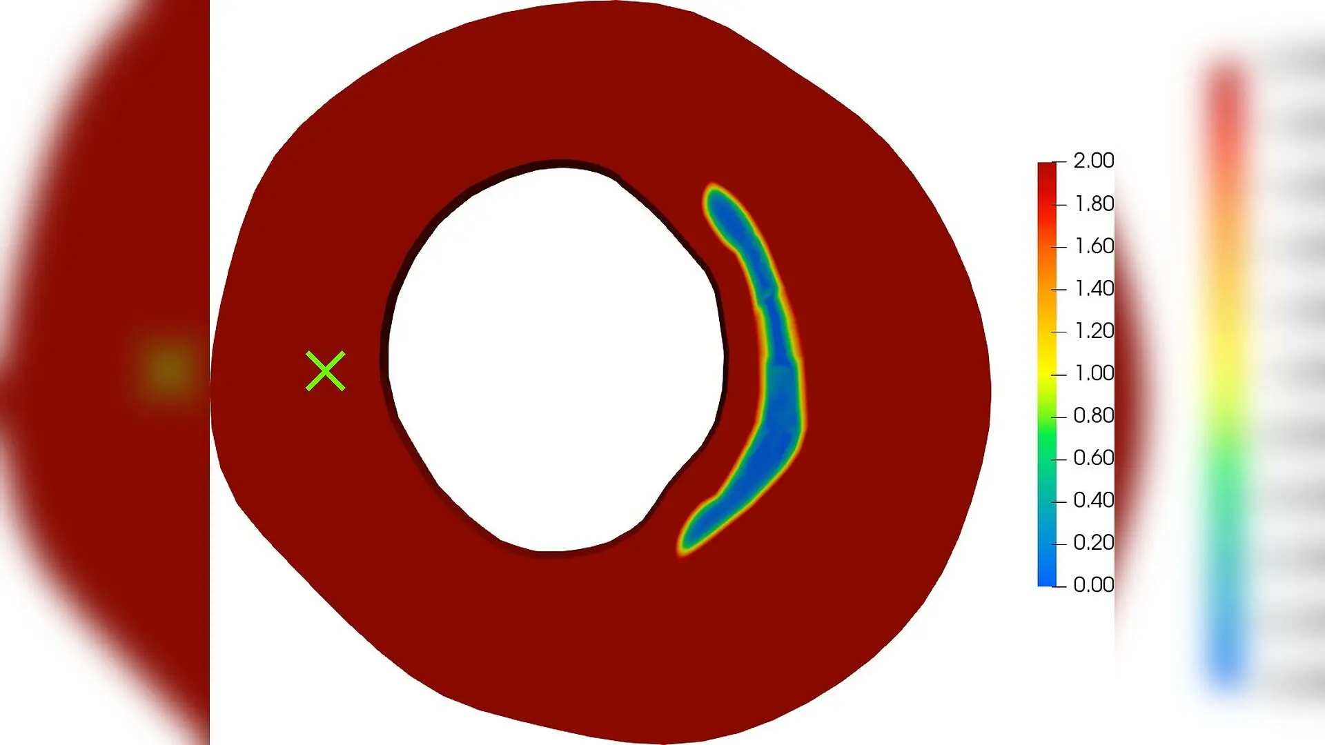
When patients take medications to lower high blood pressure, their arteries generally relax, but a new computational study reveals an unexpected mechanical side effect that could carry hidden risks for vulnerable blood vessels. The research, available on the arXiv preprint server, shows that common blood pressure drugs can unintentionally shift physical stress onto fragile protective layers within diseased arteries.
Cardiovascular disease remains the leading cause of global mortality, taking approximately 19.8 million lives in 2022. Hypertension, or high blood pressure, affects roughly 1.28 billion adults aged 30 to 79 and stands as a major driver of premature death. To combat this crisis, millions of people rely on antihypertensive medications such as amlodipine, a widely prescribed calcium channel blocker that relaxes the smooth muscle cells in blood vessel walls.
While relaxing these muscles typically reduces overall tension and lowers blood pressure, atherosclerotic arteries feature complex plaque formations containing lipid pools, calcifications, and fibrous caps. Researchers Sharan Nurani Ramesh, Lea Saßmannshausen, Axel Klawonn, and Daniel Balzani built an advanced computational model to investigate how these drugs alter local mechanical forces inside diseased arteries. Their findings indicate that as smooth muscle cells relax, they shed their load-bearing duties, forcing neighboring structural components to pick up the slack.
How Blood Pressure Drugs Shift Mechanical Loads
To understand what happens inside a medicated vessel, the team developed a mathematical framework that couples drug transport, smooth muscle cell contraction, anisotropic tissue behavior, and residual stresses. The authors modeled how amlodipine diffuses through the arterial wall, inhibiting calcium channels and reducing the ability of smooth muscle cells to contract. They tested this model on five different human arterial geometries reconstructed from histological cross-sections, representing various stages of atherosclerosis and plaque morphology.

The simulations demonstrate that while drug treatment successfully lowers stress in the medial layers of the artery, it simultaneously shifts mechanical loads directly onto the fibrous cap. In an external iliac artery simulation, the von Mises stress in the fibrous cap increased by a volume-weighted median of 28.9 kilopascals, with local peaks reaching up to 95 kilopascals. Similar stress increases occurred across multiple plaque morphologies, with the most severe jumps appearing in thin cap fibroatheromas.
Why Insufficient Pressure Drops Raise Rupture Concerns
The core danger of this load-shifting phenomenon depends heavily on how effectively the medication reduces systemic blood pressure. If a drug lowers blood pressure insufficiently, the increased mechanical burden on the fibrous cap remains unmitigated. The authors simulated blood pressure drops of 10, 20, and 30 millimeters of mercury to measure the offsetting effects of systemic relief.
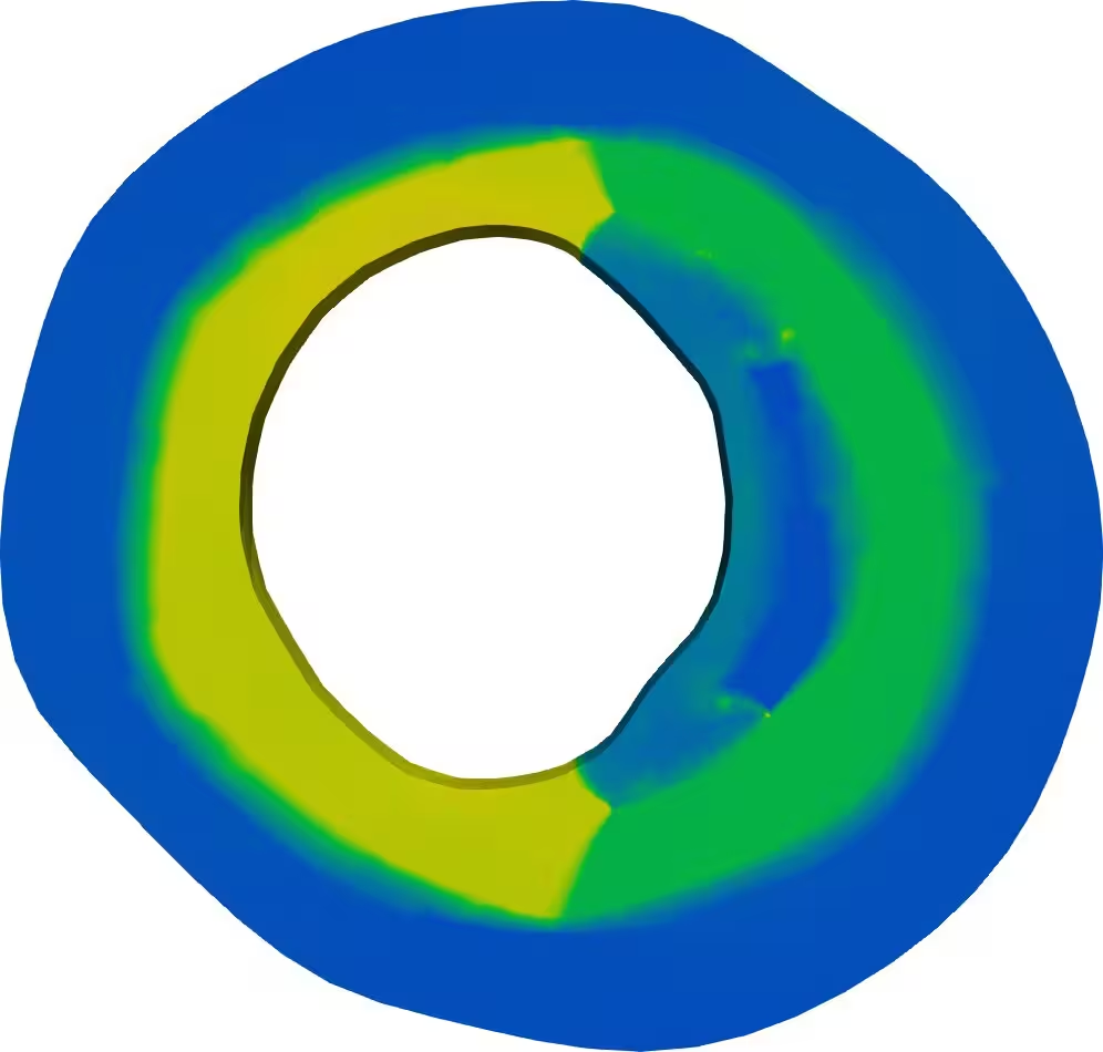
At a modest blood pressure reduction of 10 millimeters of mercury, the median stress increase in the fibrous cap dropped to 3.8 kilopascals or lower in four of the five tested geometries. When pressure drops reached 20 to 30 millimeters of mercury, the median change in stress turned neutral or negative in most arteries, meaning the benefits of lower blood pressure successfully offset the local stress increase. However, in specific geometries like the external iliac artery, moderate pressure drops still left residual stress increases of over 12 kilopascals.
What This Means for Plaque Stability and Future Treatments
An increase in fibrous cap stress does not automatically guarantee that a plaque will rupture, as the outcome depends on the intrinsic material strength of the cap itself. Human fibrous caps possess a wide strength range, spanning from 72 to 259 kilopascals. In cases where a plaque is already loaded close to its breaking point before treatment, a drug-induced stress increase could elevate the risk of rupture if blood pressure reduction is incomplete.
The researchers emphasize that major clinical trials have consistently shown long-acting calcium channel blockers to be safe and effective at reducing cardiovascular events. These large trials typically achieve sufficient blood pressure control, which aligns with the study’s finding that adequate pressure reduction neutralizes the stress shift. Nevertheless, the authors suggest that identifying patient-specific plaque vulnerabilities and ensuring optimal blood pressure control remain critical factors in designing safer therapeutic protocols.
The study was posted to the arXiv preprint server on November 16, 2026.
This article has been fact checked for accuracy, with information verified against reputable sources. Learn more about us and our editorial process.
Last reviewed on .
Article history
- Latest version
Reference(s)
- Ramesh, Sharan. “Pharmaco-mechanical computational simulation of atherosclerotic arteries: effects of calcium channel blockers on local stress distributions.” arXiv.org <http://arxiv.org/abs/2611.21320v1>.
Cite this page:
- Posted by Priya Sharma