Blood Cancer Study Links Epigenetic Protein To Survival Outcomes
Researchers find that elevated UHRF1 levels predict aggressive disease progression and poorer survival in adult T-cell leukemia/lymphoma patients.
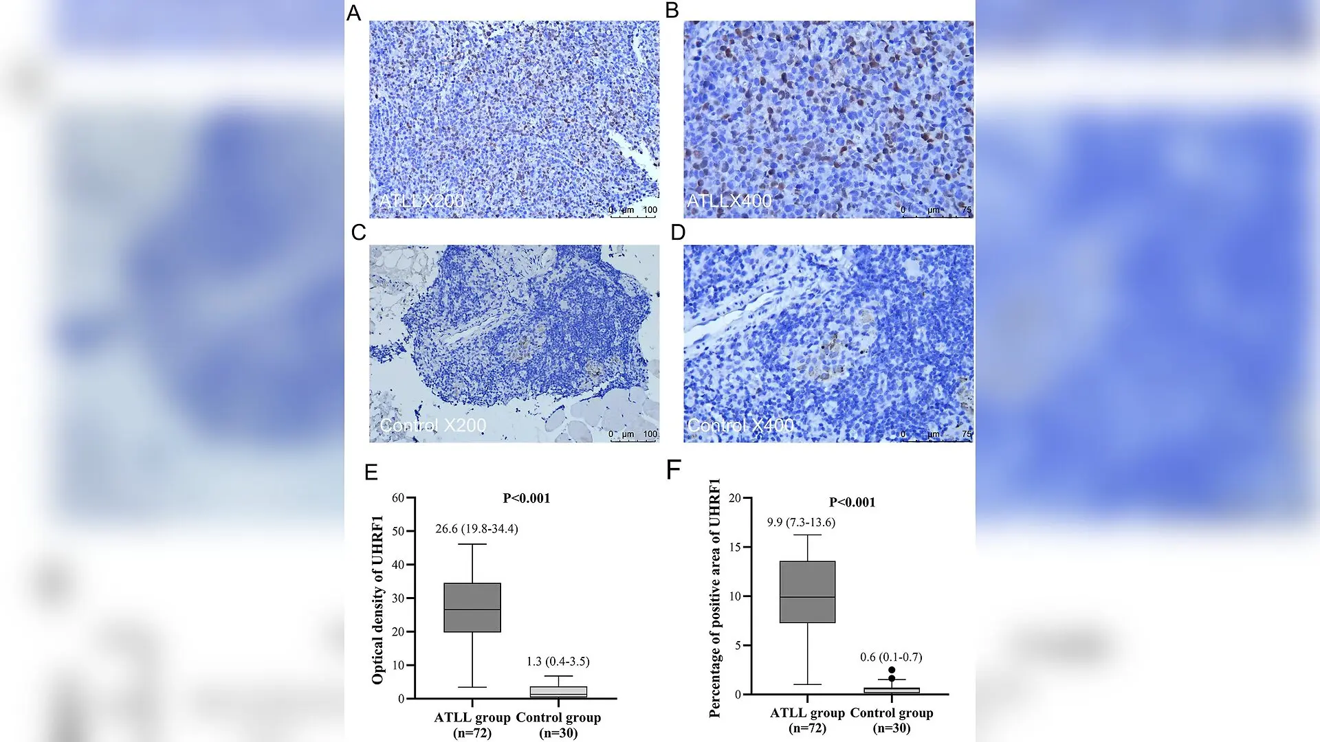
An epigenetic protein normally responsible for maintaining DNA methylation during cell division is heavily overexpressed in adults suffering from a rare and aggressive blood cancer called adult T-cell leukemia/lymphoma. Researchers discovered that measuring levels of this protein could help doctors predict which patients face the highest risk of rapid disease progression and poor survival outcomes.
Adult T-cell leukemia/lymphoma, or ATLL, is a fast-moving malignancy of mature T-lymphocytes that is driven by persistent infection with the human T-cell leukemia virus type 1, known as HTLV-1. Despite intensive therapies, patient prognoses vary widely, and clinical staging alone often fails to capture how aggressive the disease will behave in individual cases. To uncover better molecular signposts, a research team led by scientists at Fujian Medical University Union Hospital investigated the role of UHRF1, a multifunctional epigenetic regulator essential for chromatin remodeling and DNA repair.
Elevated Protein Levels in Patient Tissues
The investigators examined tumor tissue samples collected from 72 ATLL patients treated between January 2018 and January 2025, comparing them against control tissues from 30 individuals with reactive lymphadenopathy. Using immunohistochemical staining quantified via digital image analysis, the team evaluated the optical density and percentage of positive pixels for the UHRF1 protein. The results, published in PloS one, revealed that the median optical density of UHRF1 reached 26.562 in ATLL tissue compared to just 1.306 in control samples, a statistically significant difference.
Further analysis connected this overexpression to severe clinical features. High UHRF1 levels correlated significantly with acute or lymphoma-type disease subtypes, systemic B symptoms, bone marrow invasion, elevated serum lactate dehydrogenase, and higher counts of the cellular proliferation marker Ki-67. Patients with high UHRF1 expression also carried significantly higher viral loads of the underlying HTLV-1 infection.
Survival Risks and Disease Progression
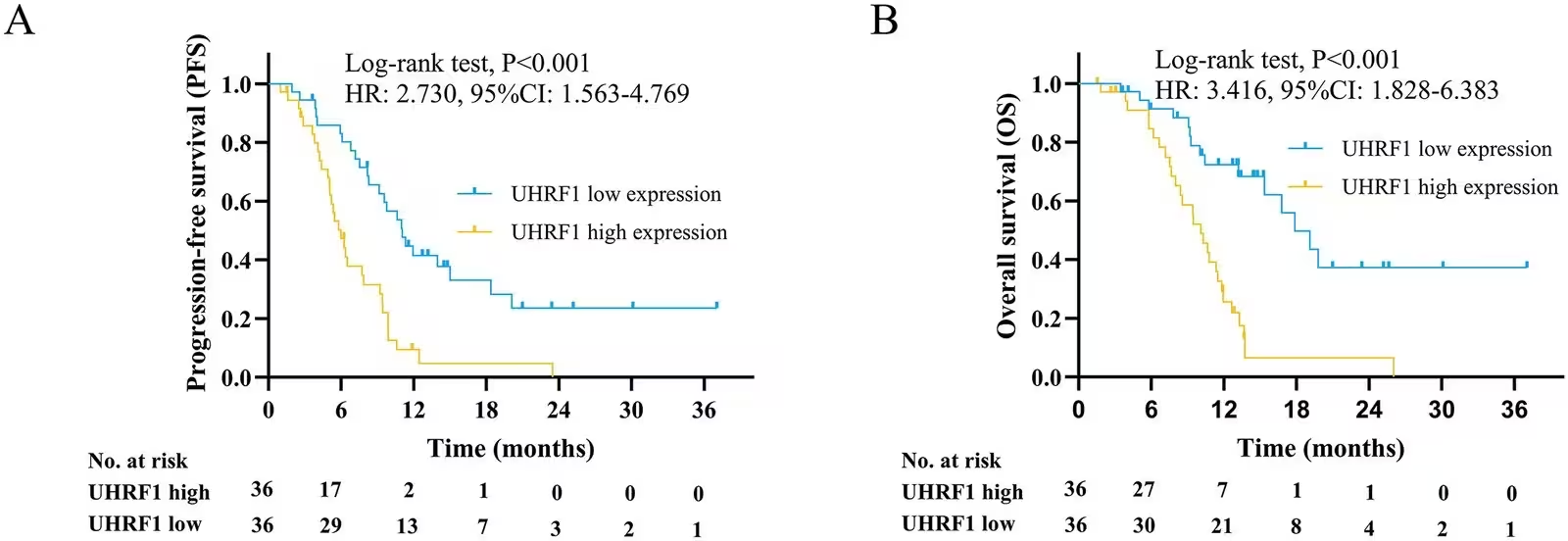
To determine the prognostic value of the protein, the researchers tracked patient survival outcomes over a median follow-up period of 10.5 months. During this window, 56 patients experienced disease progression and 43 died. Kaplan-Meier survival analysis showed that patients with elevated UHRF1 expression suffered significantly shorter progression-free survival and overall survival compared to those with low expression.
In multivariate Cox proportional hazards regression models that adjusted for confounding clinical variables, high UHRF1 expression remained an independent adverse predictor for both progression-free survival, with a hazard ratio of 1.959, and overall survival, with a hazard ratio of 2.397. These statistical models confirm that the protein carries prognostic weight independent of traditional staging indicators.
Laboratory Tests Reveal Cell Vulnerabilities
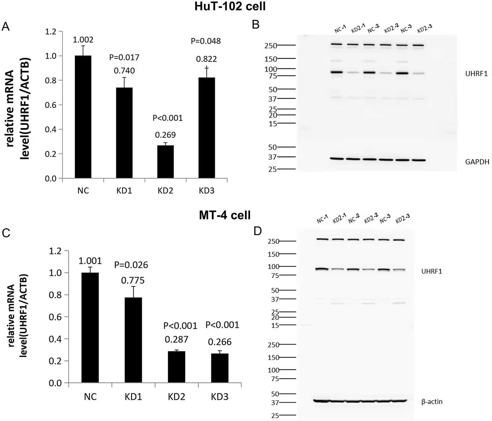
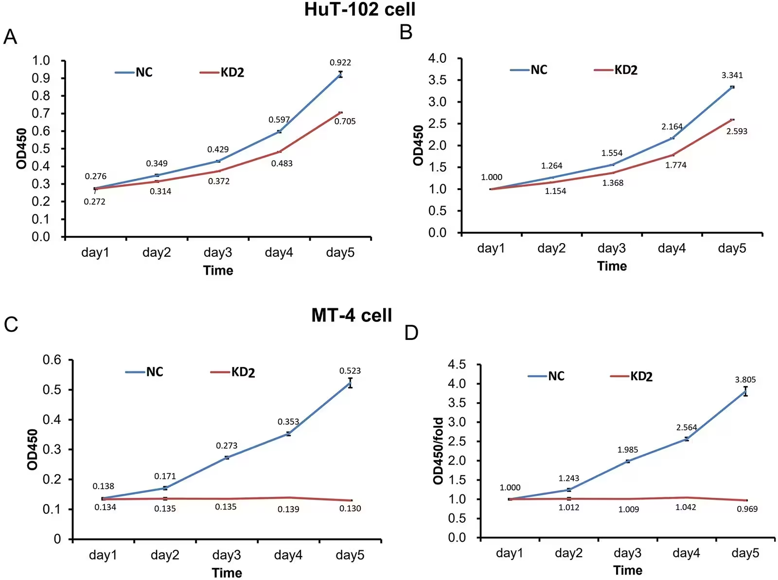
To understand what the protein does inside cancer cells, the researchers turned to laboratory experiments using human ATLL cell lines named HuT-102 and MT-4. Using short-hairpin RNA interference to silence the UHRF1 gene, the team observed dramatic changes in cell behavior. Knocking down UHRF1 significantly suppressed cell proliferation over a five-day culture period, causing severe growth arrest especially in MT-4 cells, which barely expanded at all.
Flow cytometry analysis showed that silencing the gene also triggered programmed cell death, or apoptosis. In HuT-102 cells, early and late apoptotic fractions increased significantly while viable cell percentages dropped. Furthermore, UHRF1 silencing disrupted normal cell cycle progression, prompting S-phase accumulation in HuT-102 cells and prominent G1-phase arrest in MT-4 cells.
Limits of the Current Evidence
While the experiments establish UHRF1 as a critical driver of the malignant phenotype in culture, the underlying molecular signaling pathways remain complex and incompletely mapped. The researchers investigated whether UHRF1 interacts directly with the adenosine monophosphate-activated protein kinase signaling pathway, which regulates cellular energy and metabolism. However, co-immunoprecipitation assays revealed no stable physical interaction between UHRF1 and AMPK or its regulatory phosphatase components under the tested conditions.
The authors note several limitations to their work, including its retrospective design, single-center patient cohort of 72 individuals, and reliance on cell line models. They emphasize that larger, multi-center prospective studies are needed to validate UHRF1 as a clinical biomarker and to explore its therapeutic potential in living biological models.
The research was published in PloS one on January 1, 2026.
This article has been fact checked for accuracy, with information verified against reputable sources. Learn more about us and our editorial process.
Last reviewed on .
Article history
- Latest version
Reference(s)
- Lin, Ying., et al. “Elevated UHRF1 predicts unfavorable clinical outcomes in adult T-cell leukemia/lymphoma: Clinicopathological analysis and preliminary in vitro observations.” PLOS One, vol. 21, no. 11, November 11, 2026, pp. e0362674 Public Library of Science (PLoS), doi: 10.1371/journal.pone.0362674. <https://doi.org/10.1371/journal.pone.0362674>.
Cite this page:
- Posted by Priya Sharma