Blocked Chemical Signals Alter Brain Waves During Memory Recall
Intracranial recordings reveal that disrupting acetylcholine alters hippocampal brain waves during memory retrieval even when recall performance remains completely normal.
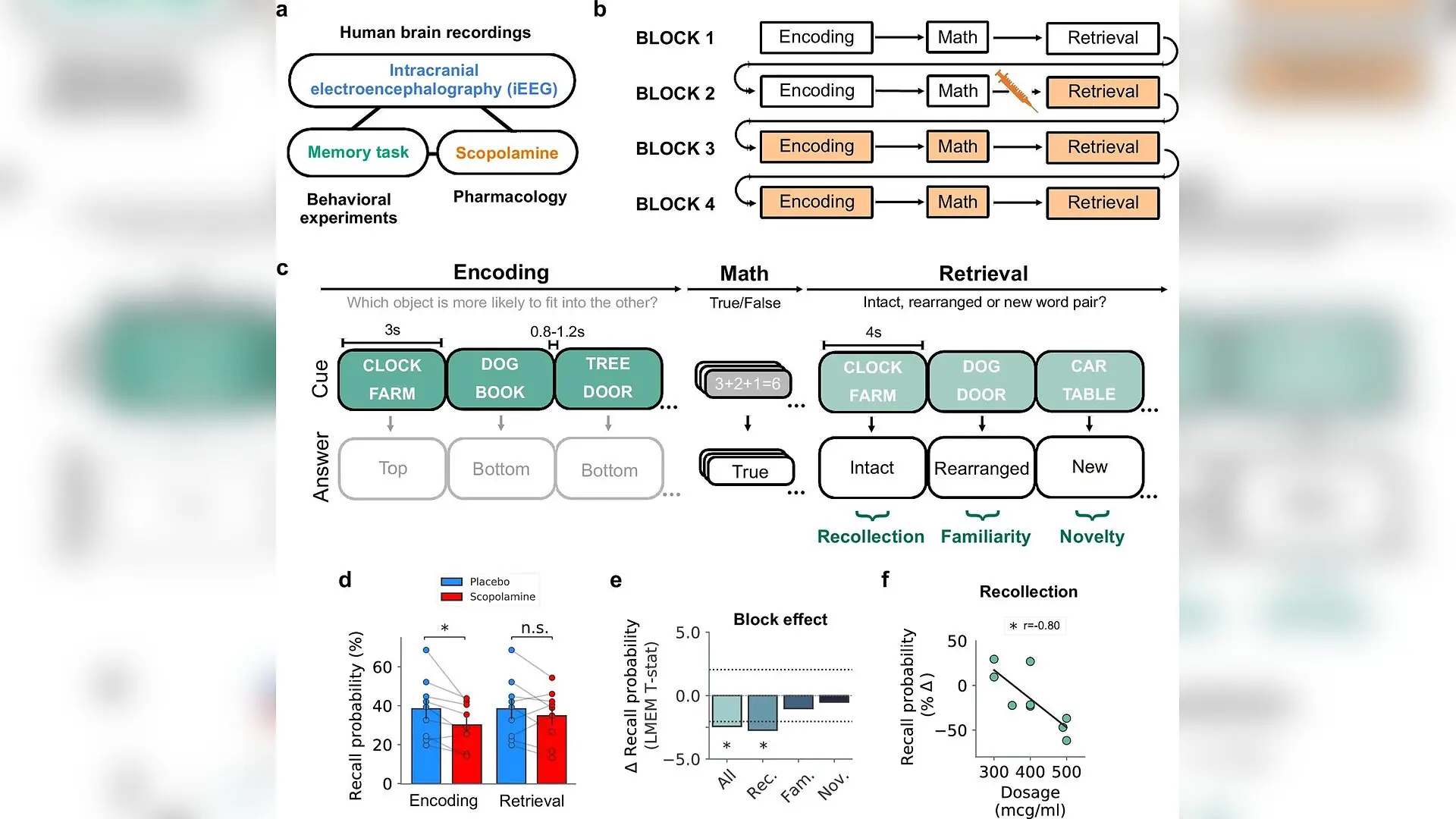
When brain chemical signals linked to attention and memory are blocked, human brain wave patterns shift during memory recall even though performance remains completely normal. Researchers investigated 12 neurosurgical epilepsy patients outfitted with intracranial depth electrodes to discover how interfering with acetylcholine, a vital neurotransmitter, changes the electrical activity of the hippocampus during memory tasks.
Loss of cholinergic signaling is a classic hallmark of Alzheimer’s disease and other severe cognitive disorders. Yet, the exact neurophysiological mechanisms connecting these chemical imbalances to human memory formation and retrieval have remained poorly understood. To isolate these effects, Tamara Gedankien and colleagues administered scopolamine, a muscarinic cholinergic receptor antagonist, in a double-blind, within-subject design as patients performed an associative recognition memory task involving word pairs. The study is published in eLife, detailing how intracranial electroencephalography captured brain dynamics across four experimental blocks.
How Drug Blockade Separates Memory Encoding from Retrieval
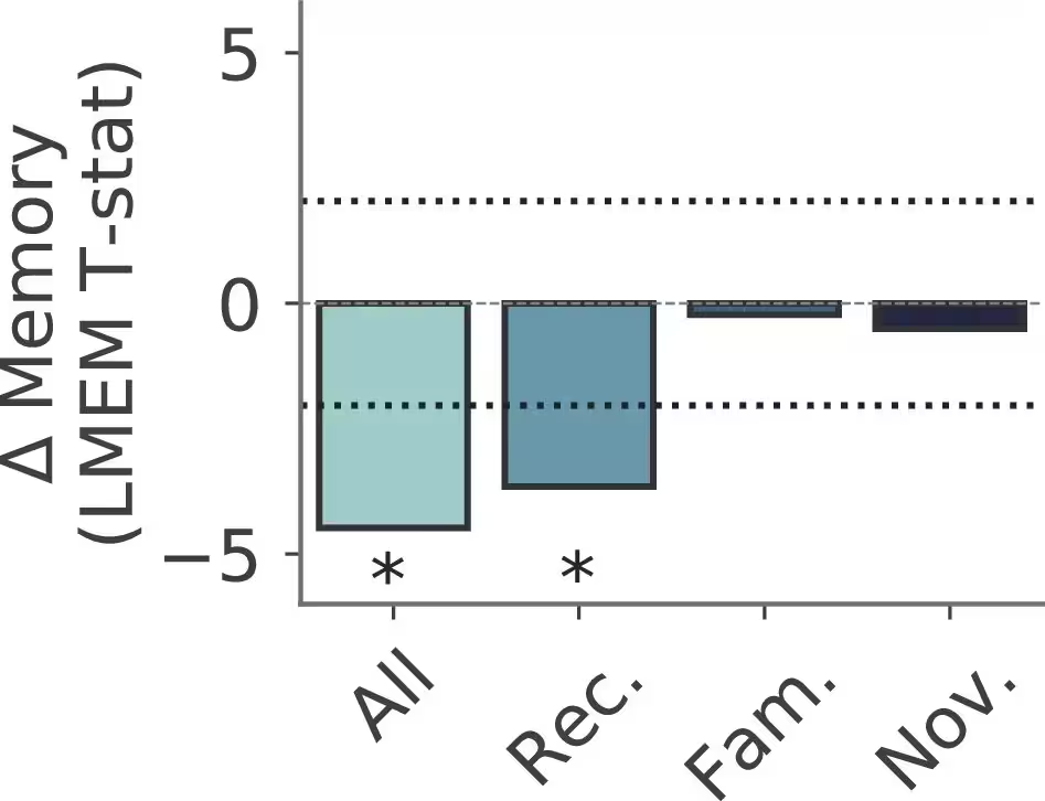
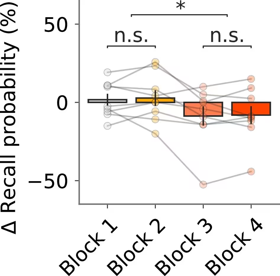
During the experiments, participants completed tasks under baseline conditions, following placebo saline injections, and under the influence of intravenous scopolamine given either prior to encoding or restricted exclusively to the retrieval stage. The team replicated earlier behavioral findings showing that when the drug was present only during the retrieval phase, memory performance stayed entirely unaffected. Conversely, administering the drug prior to encoding led to significant memory impairments that specifically targeted recollection-based memory while sparing familiarity and novelty detection.
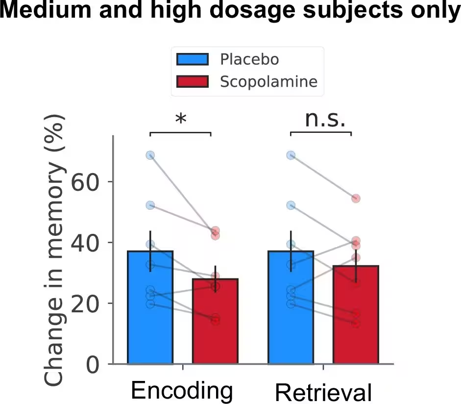
This dissociation provided a rare window to examine the hidden electrical changes occurring inside the brain during recall. Even though memory behavior stayed intact when scopolamine was given before retrieval alone, the intracranial recordings revealed significant disruptions. Slow theta oscillations, rhythmic brain waves operating at 2 to 4 hertz, experienced dose-dependent power reductions, impaired phase reset, and a failure to reinstate encoding-related spectral patterns.
Challenging Long-Standing Theories on Brain Waves
These surprising electrical disruptions challenge the classical view that slow theta oscillations are strictly necessary for memory retrieval. Scientists previously assumed that theta rhythms acted as an absolute requirement for pulling stored memories back into conscious awareness. Because theta power and phase alignment dropped significantly during retrieval under scopolamine without harming test scores, the authors suggest that theta activity appearing during recall reflects something else entirely.
Instead of passive memory playback, theta oscillations during retrieval likely serve an active role in memory updating, contextual integration, and re-encoding. The brain appears to re-engage an encoding-like neural state during retrieval to weave new contextual information into existing memories, a process that relies heavily on healthy cholinergic tone.
Decoding Neural States Through Advanced Analytics
To make sense of complex multi-frequency signals across various brain subregions, the team applied principal component analysis and machine learning classifiers to the intracranial data. Their unsupervised models revealed that nearly 80 percent of the total variance across trial recordings could be captured by a single principal component. This low-dimensional structure proved that large-scale spectral fluctuations are highly coordinated across electrodes in the hippocampus and entorhinal cortex.
Frequency-band-specific decoders demonstrated that slow theta activity carried the strongest signature for distinguishing drug states from placebo conditions. Furthermore, classification accuracy scaled positively with the number of recording electrodes included, proving that hippocampal subregions contribute distinct electrophysiological features when processing pharmacological disruption.
Limitations and Future Clinical Directions
While these findings offer unprecedented resolution into human medial temporal lobe dynamics, the study carries inherent limitations. The participant pool consisted exclusively of treatment-resistant epilepsy patients undergoing invasive monitoring, meaning their local neural circuitry may differ from healthy populations. Additionally, intravenous scopolamine acts globally on muscarinic receptors throughout the central nervous system rather than targeting specific memory circuits in isolation.
Despite these constraints, clarifying how acetylcholine shapes rhythmic brain biomarkers paves the way for future therapeutic strategies. Understanding the precise electrical fingerprints of cholinergic dysfunction could eventually inform targeted treatments for Alzheimer’s disease and related memory disorders.
The research was published in eLife on November 11, 2026.
This article has been fact checked for accuracy, with information verified against reputable sources. Learn more about us and our editorial process.
Last reviewed on .
Article history
- Latest version
Reference(s)
- Gedankien, Tamara., et al. “Cholinergic blockade reveals a role for human hippocampal theta in memory encoding but not retrieval.” eLife, vol. 14, November 11, 2026 eLife Sciences Publications, Ltd, doi: 10.7554/eLife.108972. <https://doi.org/10.7554/eLife.108972>.
Cite this page:
- Posted by Robert Brown