Engineered Nanofibrous Scaffolds Mimic Knee Cartilage Using Machine Learning
Piezoelectric scaffolds with tailored fiber architectures guide stem cell organization and predict tissue regeneration potential.
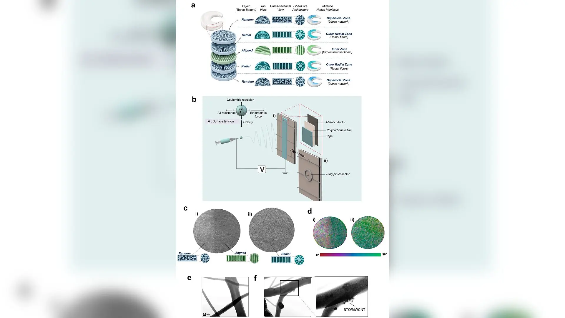
A team of researchers has developed custom electrospun nanofibrous scaffolds designed to replicate the complex structural environment of the human knee meniscus, using machine learning to map how stem cells respond to these artificial tissues. The meniscus is a crescent-shaped wedge of cartilage that absorbs shock and stabilizes the knee joint, but it heals poorly after injury due to its complex, directionally organized internal fibers. The study, published in the Journal of functional biomaterials published in the Journal of functional biomaterials, investigates how combining specific fiber arrangements with piezoelectric materials can guide cell growth.
How Custom Collector Geometries Shape Artificial Cartilage
To mimic the native meniscus, the team fabricated scaffolds using poly(vinylidene fluoride), a piezoelectric polymer that generates localized electrical signals when mechanically deformed. They incorporated barium titanate nanoparticles and multi-walled carbon nanotubes into the polymer matrix to modify its electrical and structural properties. By altering the collector setup during the electrospinning process, the researchers produced scaffolds with distinct architectures, including randomly oriented fibers, uniaxially aligned bundles, and radially organized pathways. Scanning electron microscopy confirmed that these modifications successfully directed fiber deposition without altering the underlying chemistry of the material.
What Happens When Stem Cells Meet Engineered Scaffolds
Human bone marrow-derived mesenchymal stem cells were seeded onto the scaffolds and cultured to evaluate how different topographical cues influenced cellular behavior. Quantitative fluorescence imaging revealed that aligned and radial architectures directed cellular elongation and cytoskeletal organization along specific trajectories through contact guidance. Cells cultured on the aligned scaffold formulation exhibited robust cellularity and directional alignment, while radial configurations successfully guided cells along diverging spatial pathways. The researchers also measured the expression of SOX9, a key transcription factor associated with chondrogenic activity, demonstrating that the engineered microenvironments supported phenotype-associated molecular responses alongside morphological alignment.
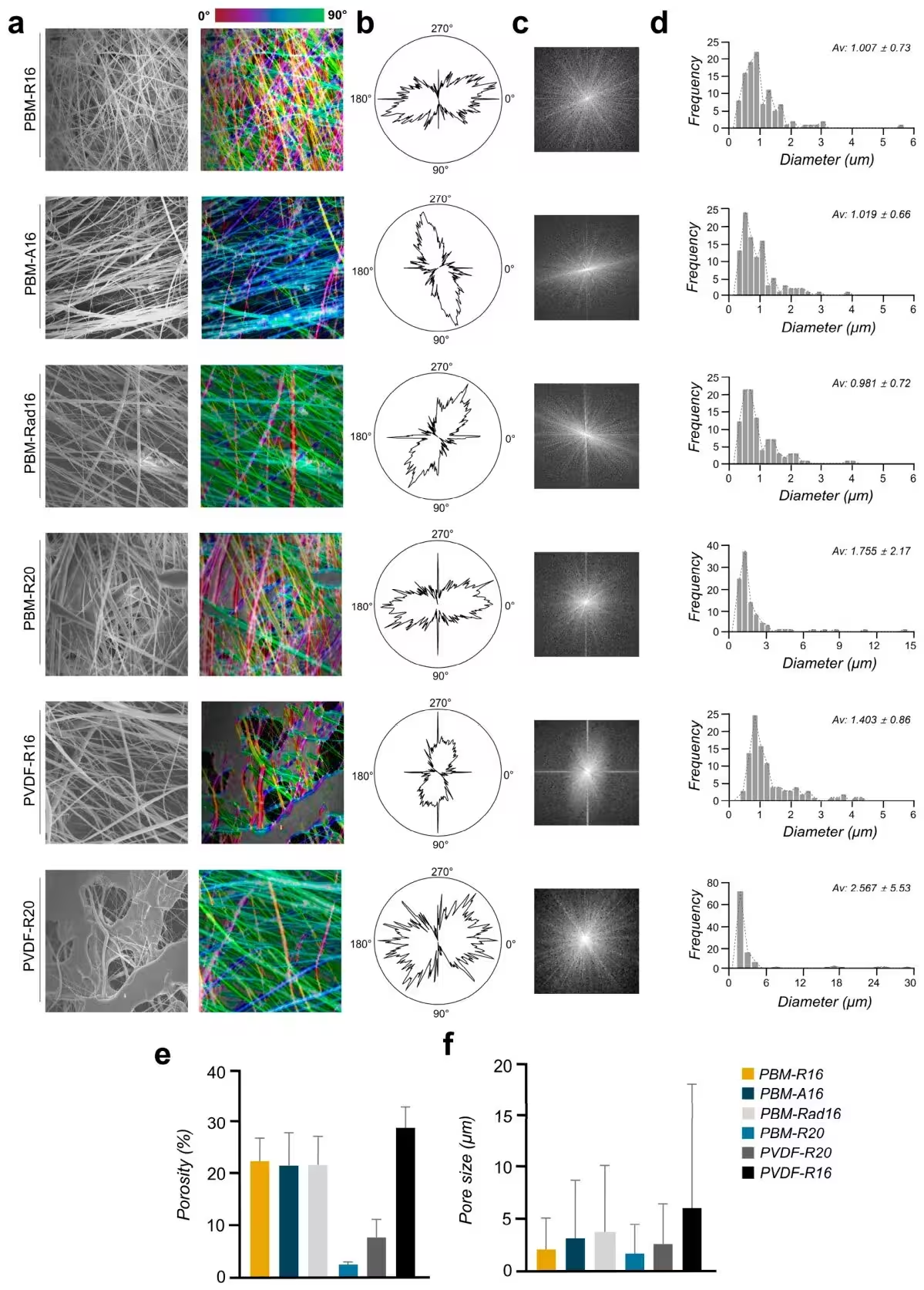
Combining Multidimensional Data With Machine Learning Models
Because individual biological markers fail to capture the full scope of cellular adaptation, the team integrated nuclear morphology, cytoskeletal organization, and SOX9 expression into a composite metric known as the Meniscus-Mimetic Scaffold Quality Index. They then fed these multidimensional parameters into a series of regression algorithms and a specialized machine learning framework to predict scaffold performance. The advanced model achieved a cross-validated R-squared of 0.853, significantly outperforming conventional linear regression and individual neural network baselines. Feature importance analysis identified the nuclear SOX9 intensity ratio and nuclear area as major contributors to the predictive accuracy of the model.
Limitations and Future Directions for Scaffold Engineering
The authors emphasize that their predictive framework remains exploratory because it is based on a limited set of scaffold formulations and experimental replicates. While the machine learning models successfully mapped relationships within the current dataset, external validation using broader formulations and longer-term mechanical testing will be necessary before these techniques can be applied to clinical tissue engineering. Future work will also need to examine how these piezoelectric scaffolds perform under dynamic mechanical loading inside a functional joint environment.
The research was published in Journal of functional biomaterials on October 21, 2026.
This article has been fact checked for accuracy, with information verified against reputable sources. Learn more about us and our editorial process.
Last reviewed on .
Article history
- Latest version
Reference(s)
- Kim, Jeong In., et al. “Machine Learning-Assisted Evaluation of Topography-Dependent Cellular Responses in Meniscus-Mimetic Piezoelectric Nanofibrous Scaffolds.” Journal of Functional Biomaterials, vol. 17, no. 10, October 21, 2026, pp. 532 MDPI AG, doi: 10.3390/jfb17100532. <https://doi.org/10.3390/jfb17100532>.
Cite this page:
- Posted by Robert Brown