AI Enhances Label-Free Optical Microscopy Past Century-Old Diffraction Limit
Custom deep learning architectures O-Net and Θ-Net resolve nanoscale features down to 170 nanometers without fluorescent labels, depending on input image quality.
Researchers have developed artificial intelligence models that push label-free optical microscopy beyond its classical limits, successfully recovering nanoscale features as small as 170 nanometers using phase-dependent imaging techniques. Optical microscopy has remained constrained for more than a century by the Rayleigh criterion, a fundamental diffraction barrier that prevents standard lenses from resolving details smaller than roughly 200 nanometers under visible light. While advanced fluorescence nanoscopy techniques bypass this restriction, they require chemical labels that frequently cause phototoxicity, quenching, and sample perturbation. To solve this limitation without exogenous markers, scientists are increasingly turning to computational super-resolution approaches that enhance conventional micrographs using deep neural networks.
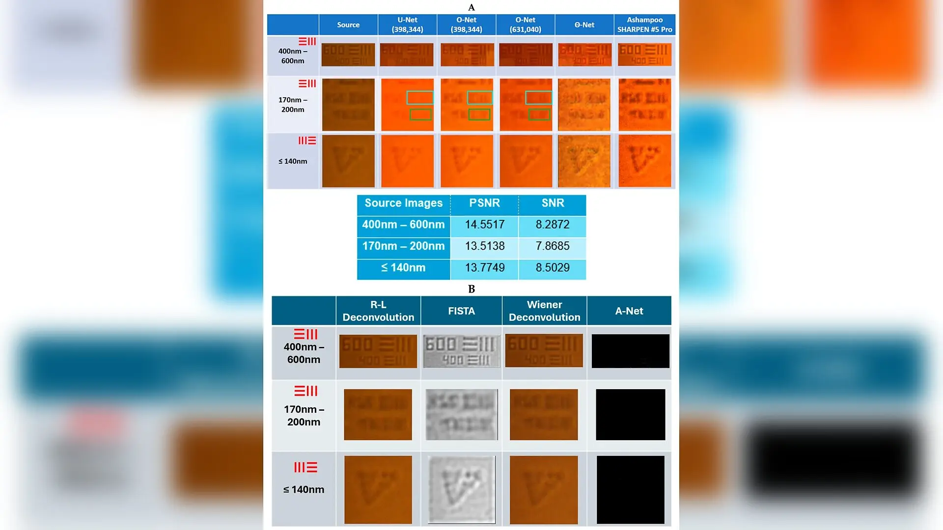
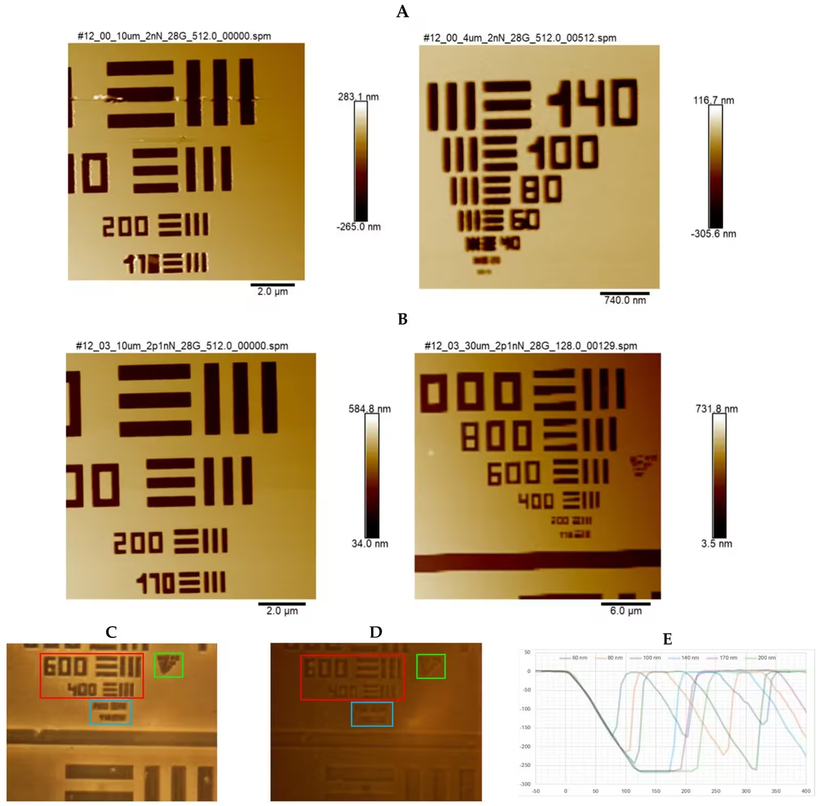
In a study published in Nanomaterials, a team of researchers systematically evaluated two custom deep learning architectures named O-Net and Θ-Net against standard U-Net models for label-free phase-dependent microscopy. The study utilized quantitative phase imaging techniques, specifically phase-contrast microscopy and differential interference contrast microscopy, which generate image contrast from intrinsic refractive index variations rather than fluorescent dyes. Because learning-based restoration can sometimes invent non-existent structures or hallucinate high-frequency details, the team rigorously validated their computer models using a custom-fabricated nanoscale test target calibrated with atomic force microscopy. This physical reference standard featured precision-etched bars and spaces ranging from 20 nanometers to 6 micrometers, allowing the team to measure actual structural recovery rather than relying on visual enhancement alone.
How Signal Quality Dictates Neural Network Success
The investigation revealed that the effectiveness of a specific neural network architecture depends heavily on the signal-to-noise ratio of the input image. Under moderately high signal-to-noise ratio conditions achieved at an intermediate magnification of 2.4 times, the single-node O-Net architecture produced the cleanest reconstructions with clear line delineation and lower peak error metrics. Specifically, the O-Net models successfully resolved 200-nanometer features in differential interference contrast images and 170-nanometer features in phase-contrast images, representing resolution enhancement factors of up to 1.94 times beyond the calculated theoretical diffraction limit of approximately 325 to 329 nanometers.
However, when the optical magnification was increased to 3.75 times, the input image signal-to-noise ratio dropped significantly due to reduced light collection, making it difficult to distinguish fine bars from background noise. Under these challenging low-signal conditions, the multi-node Θ-Net architecture outperformed both O-Net and U-Net models by remaining sensitive to subtle pixel intensity fluctuations. The researchers identified empirical transition thresholds around a signal-to-noise ratio of 7.5 for differential interference contrast and 11 for phase-contrast microscopy, where lower values favored Θ-Net and higher values favored O-Net.
Understanding the Limits of Computational Nanoscopy
To ensure that the enhanced images represented genuine physical recovery rather than algorithmic artifacts, the team analyzed intensity profile plots across the reconstructed features and evaluated contrast-to-noise ratios, contrast transfer functions, and full-width-at-half-maximum measurements. Traditional image deconvolution algorithms such as Richardson-Lucy, Wiener filtering, and the fast iterative shrinkage-thresholding algorithm struggled to resolve the nanoscale features because low input contrast hindered their ability to separate signal from noise. The deep learning models succeeded by operating across both spatial and Fourier frequency domains, applying learned deconvolution kernels that effectively extended the sampling limits of the optical train.
Despite these promising results, the authors emphasize important technical caveats. Enhanced contrast or sharper boundaries do not automatically prove that hidden spatial information has been perfectly recovered, and features approaching 140 nanometers remained largely unresolvable or prone to ambiguity. Furthermore, because these neural networks learn from specific optical configurations, transferring the models to a different microscope or camera setup would require complete retraining. Future efforts will focus on incorporating polarization variables for differential interference contrast and developing universally standardized image quality metrics for label-free microscopy.
The research was published in Nanomaterials (Basel, Switzerland) on October 21, 2026.
This article has been fact checked for accuracy, with information verified against reputable sources. Learn more about us and our editorial process.
Last reviewed on .
Article history
- Latest version
Reference(s)
- Kaderuppan, Shiraz S.., et al. “Empirical Validation of SNR-Dependent Deep Learning Architectures for Label-Free Super-Resolution in Optical Microscopy.” Nanomaterials, vol. 16, no. 20, October 21, 2026, pp. 1334 MDPI AG, doi: 10.3390/nano16201334. <https://doi.org/10.3390/nano16201334>.
Cite this page:
- Posted by Robert Brown