Embryonic Heat Waves Leave Permanent Scars on Developing Fly Brains
Biotechnology

Embryonic Heat Waves Leave Permanent Scars on Developing Fly Brains

Brief thermal stress during a narrow embryonic window alters central neural circuits and slows larval movement while peripheral synapses successfully compensate.
By Robert Brown
Published:
Email this Article
Embryonic Heat Waves Leave Permanent Scars on Developing Fly Brains
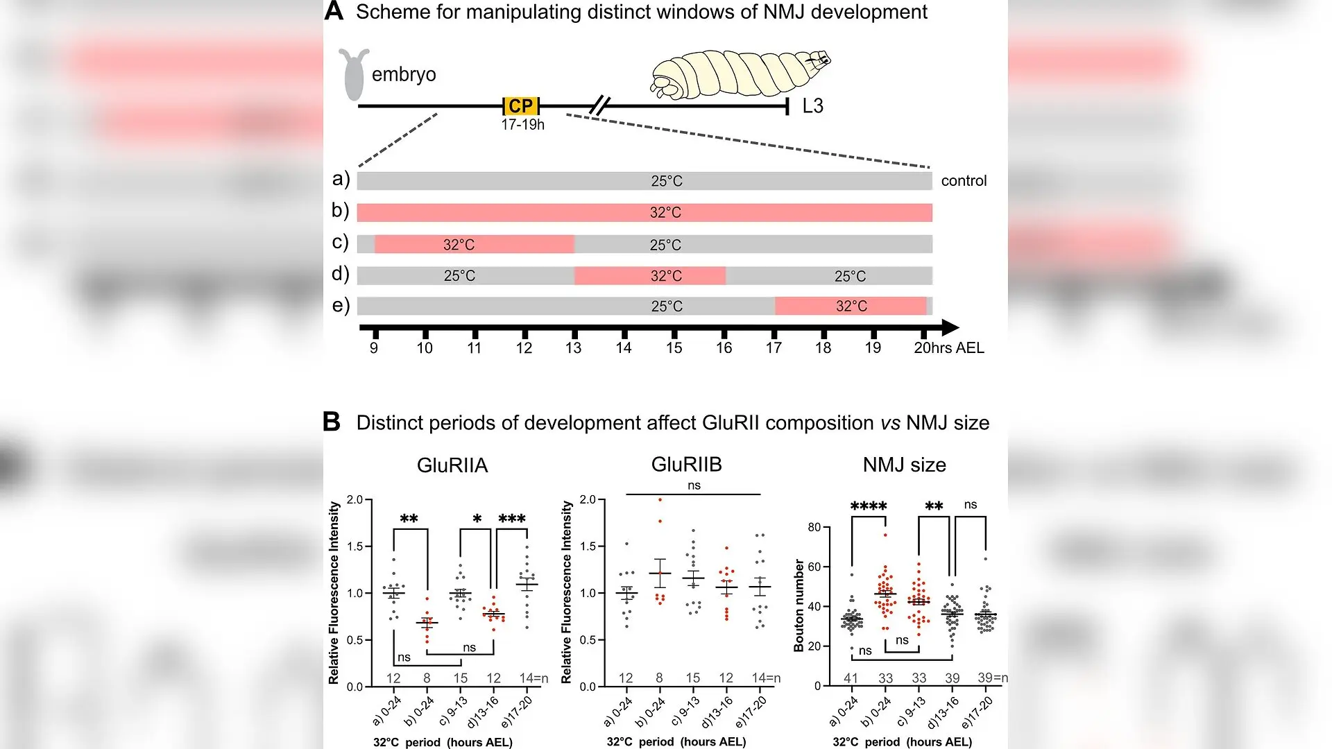
Heat stress perturbations during distinct periods of development differentially affect postsynaptic GluRII receptor composition vs neuromuscular junction (NMJ) size. (A) Schematic of temperature manipulations during embryogenesis. Embryos collected over 1 hr were incubated at 32 °C for different phases of embryogenesis. (B) Resultant changes to the development of the aCC NMJ on muscle dorsal acute muscle 1 (DA1) were quantified at the late wandering third instar larval stage. Reduced expression of the GluRIIA subunit occurred following heat stress experienced during 13–16 hr after egg laying (AEL), comparable to GluRIIA changes caused by heat stress during all of embryogenesis, but not in response to heat stress experienced before or after this 3 hr window. GluRIIB levels are not affected by heat stress during any period or all of embryogenesis. NMJ size, in contrast, is affected by heat stress experienced during earlier phases of muscle development, from 9 to 13 hr AEL. Data are shown Niklas Krick et al. / eLife

When developing fruit fly embryos experience a brief heat wave, their nervous systems undergo lasting structural shifts that impair coordination and reduce network stability long after hatching. The research, published in eLife, reveals how different parts of a simple motor circuit respond in entirely different ways to the exact same thermal stress during early development.

During nervous system development, young organisms pass through transient windows of heightened plasticity known as critical periods. Perturbations occurring inside these specific developmental windows can trigger permanent structural and functional changes in neural networks, whereas identical disturbances outside those windows pass without consequence. While neuroscientists have long studied critical periods in mammalian brains and avian imprinting, the precise cellular mechanisms remain difficult to untangle in complex mammalian cortices.

To overcome this experimental barrier, a team of researchers led by scientists at the University of Cambridge and the University of Manchester turned to the larval locomotor network of the fruit fly, Drosophila melanogaster. Equipped with a fully mapped neural connectome and advanced genetic tools, the team set out to investigate how distinct, connected cellular nodes within a defined motor circuit adapt or maladapt when exposed to environmental stress.

How Thermal Stress Disrupts Developing Motor Networks

As poikilothermic animals whose internal temperatures match their surroundings, developing fruit flies are highly sensitive to thermal fluctuations. Previous studies established that a critical period for locomotor network development occurs late in embryogenesis, specifically between 17 and 19 hours after egg laying. The researchers tested the effects of transiently exposing embryos to a 32 degrees Celsius heat stress, which is seven degrees above the standard laboratory rearing temperature of 25 degrees Celsius but well within the range of temperatures these insects encounter in the wild.
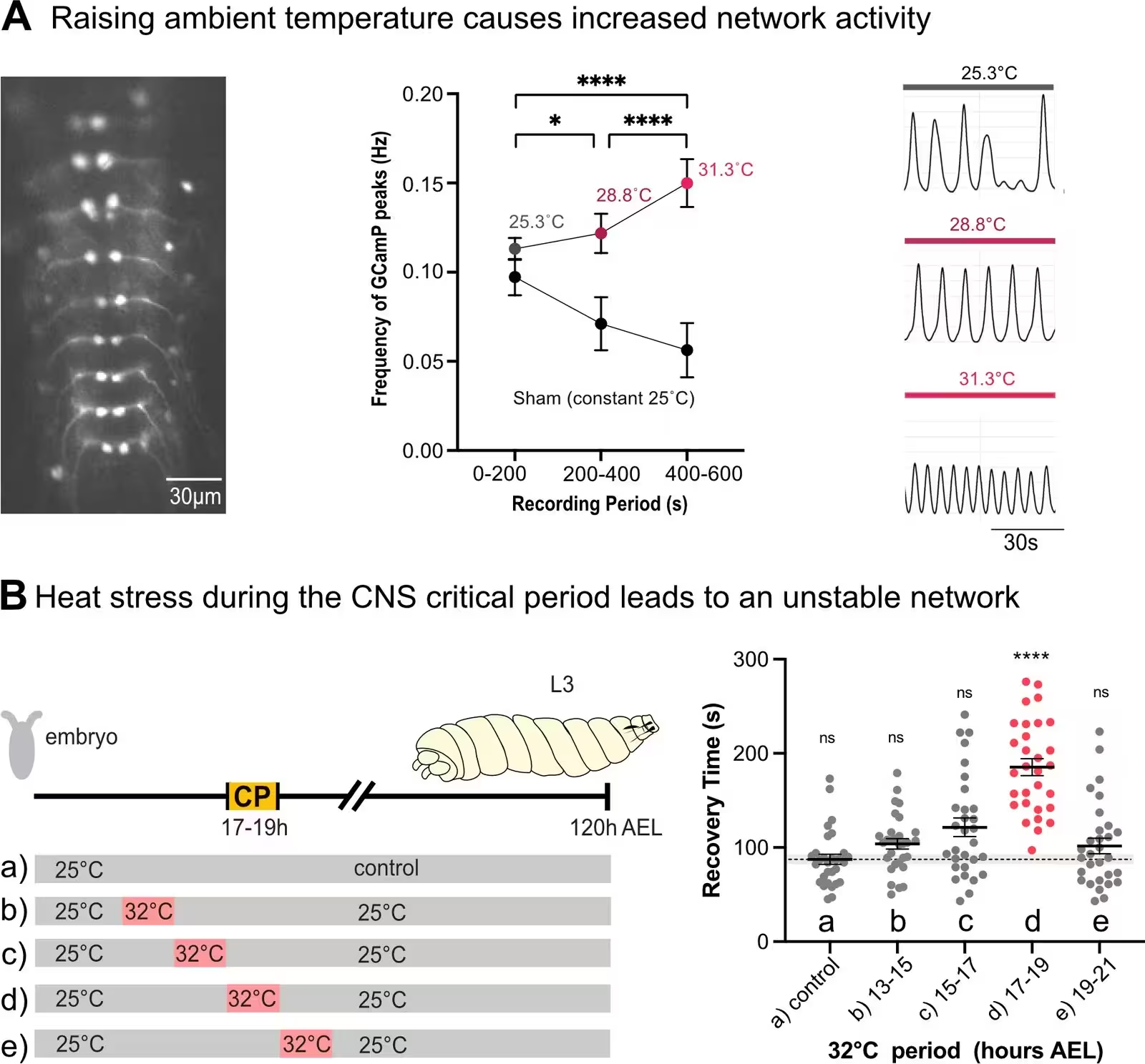
Figure 1: Transient heat stress during the critical period leads to an unstable network. (A) Acute increase of ambient temperature leads to increased network activity, as measured with the fluorescent calcium indicator GCaMP8f expressed in segmentally repeated aCC motoneurons in isolated central nervous systems; while constant 25 °C leads to reduced activity over time. Left: micrograph of an isolated ventral nerve cord with segmentally repeated aCC motoneurons expressing GCaMP8f. Centre: traces of GCaMP fluorescence peaks measured at different temperatures. Right: quantification of fluorescence peak frequency changes over time (control/sham - bottom trace, n=10 nerve cords) and in response to temperature increase (top trace, n=10 nerve cords). (B) Heat stress, when experienced during the embryonic critical period (CP) of locomotor network development, leads to reduced network stability. Left: Schematic representation of embryonic and larval development and the paradigm of temperature manipulatio
Transient heat stress during the critical period leads to an unstable network. (A) Acute increase of ambient temperature leads to increased network activity, as measured with the fluorescent calcium indicator GCaMP8f expressed in segmentally repeated aCC motoneurons in isolated central nervous systems; while constant 25 °C leads to reduced activity over time. Left: micrograph of an isolated ventral nerve cord with segmentally repeated aCC motoneurons expressing GCaMP8f. Centre: traces of GCaMP fluorescence peaks measured at different temperatures. Right: quantification of fluorescence peak frequency changes over time (control/sham - bottom trace, n=10 nerve cords) and in response to temperature increase (top trace, n=10 nerve cords). (B) Heat stress, when experienced during the embryonic critical period (CP) of locomotor network development, leads to reduced network stability. Left: Schematic representation of embryonic and larval development and the paradigm of temperature manipulatio Credit: Niklas Krick et al. / eLife

When embryos experienced this two-hour heat stress outside the critical period, either earlier or later in gestation, the resulting third-instar larvae developed normal neurological function. However, when the heat stress coincided precisely with the 17 to 19 hour critical period, the larvae hatched with reduced crawling speeds and diminished network stability. When subjected to electroshock-induced seizures later in life, these heat-stressed animals took significantly longer to recover than controls, indicating an underlying vulnerability in their central nervous circuitry.

Distinct Cellular Nodes Follow a Functional Hierarchy

To pinpoint which components of the nervous system drove these behavioral deficits, the authors examined individual connected elements within the motor circuit, ranging from central interneurons to motoneurons and peripheral neuromuscular junctions. They discovered that each cell type reacted to embryonic heat stress through vastly different mechanisms.
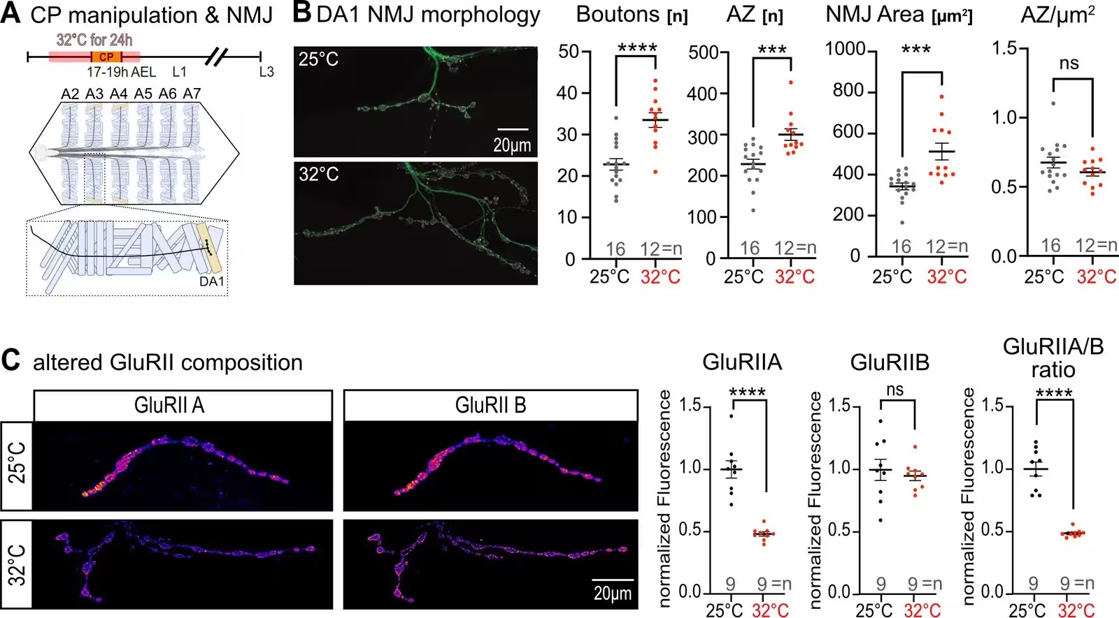
Figure 2: Morphological changes at the neuromuscular junction after critical period perturbation. (A) Schematic of temperature manipulation during embryogenesis and location of dorsal acute muscle 1 (DA1) within a fillet dissected larva. Embryos collected over a 6 hr period were incubated at 32 °C for 24 hr to guarantee heat stress exposure during the locomotor network critical period (CP). (B) This results in a change of subsequent neuromuscular junction (NMJ) development, which manifests as a significant overgrowth of aCC presynaptic terminals (green, visualised with anti-HRP-AlexaFluor488). The increase in bouton number and NMJ area is paralleled by concomitant increases in active zones (magenta), resulting in a normal active zone density. (C) Postsynaptically, embryonic heat stress causes a significant decrease of glutamate receptors containing the larger conductance GluRIIA subunit, while those with the GluRIIB subunit remain unaffected. Thus, the postsynaptic GluRII receptor composition is
Morphological changes at the neuromuscular junction after critical period perturbation. (A) Schematic of temperature manipulation during embryogenesis and location of dorsal acute muscle 1 (DA1) within a fillet dissected larva. Embryos collected over a 6 hr period were incubated at 32 °C for 24 hr to guarantee heat stress exposure during the locomotor network critical period (CP). (B) This results in a change of subsequent neuromuscular junction (NMJ) development, which manifests as a significant overgrowth of aCC presynaptic terminals (green, visualised with anti-HRP-AlexaFluor488). The increase in bouton number and NMJ area is paralleled by concomitant increases in active zones (magenta), resulting in a normal active zone density. (C) Postsynaptically, embryonic heat stress causes a significant decrease of glutamate receptors containing the larger conductance GluRIIA subunit, while those with the GluRIIB subunit remain unaffected. Thus, the postsynaptic GluRII receptor composition is Credit: Niklas Krick et al. / eLife

Within the central nervous system, transient heat stress triggered an increase in excitatory synaptic drive from premotor interneurons, such as the A27h interneuron, onto downstream motoneurons. Interestingly, the postsynaptic motoneurons appeared to compensate for this excess excitation by reducing their own intrinsic excitability. This homeostatic adjustment successfully preserved normal firing patterns at the level of individual motoneurons and muscle fibers, yet the overall propagation speed of neuronal activation waves across abdominal segments remained significantly slowed.

Peripheral Synapses Maintain Normal Transmission Despite Overgrowth

At the periphery, the neuromuscular junction connecting motoneurons to body wall muscles showed striking structural alterations following embryonic heat stress. Presynaptic terminals exhibited significant overgrowth, featuring roughly a 30 percent increase in active zones and a higher bouton count. Simultaneously, the postsynaptic receptor composition shifted, marked by a 50 percent reduction in glutamate receptors containing the larger conductance GluRIIA subunit.

Despite these dramatic morphological changes, baseline synaptic transmission at the neuromuscular junction remained completely normal. Electrophysiological recordings using two-electrode voltage clamps demonstrated that single-pulse transmission, quantal size, and the readily releasable pool of synaptic vesicles were indistinguishable from control animals. Furthermore, the peripheral synapse successfully mounted presynaptic homeostatic potentiation when challenged with pharmacological blockers, indicating that structural remodeling does not automatically impair synaptic function.
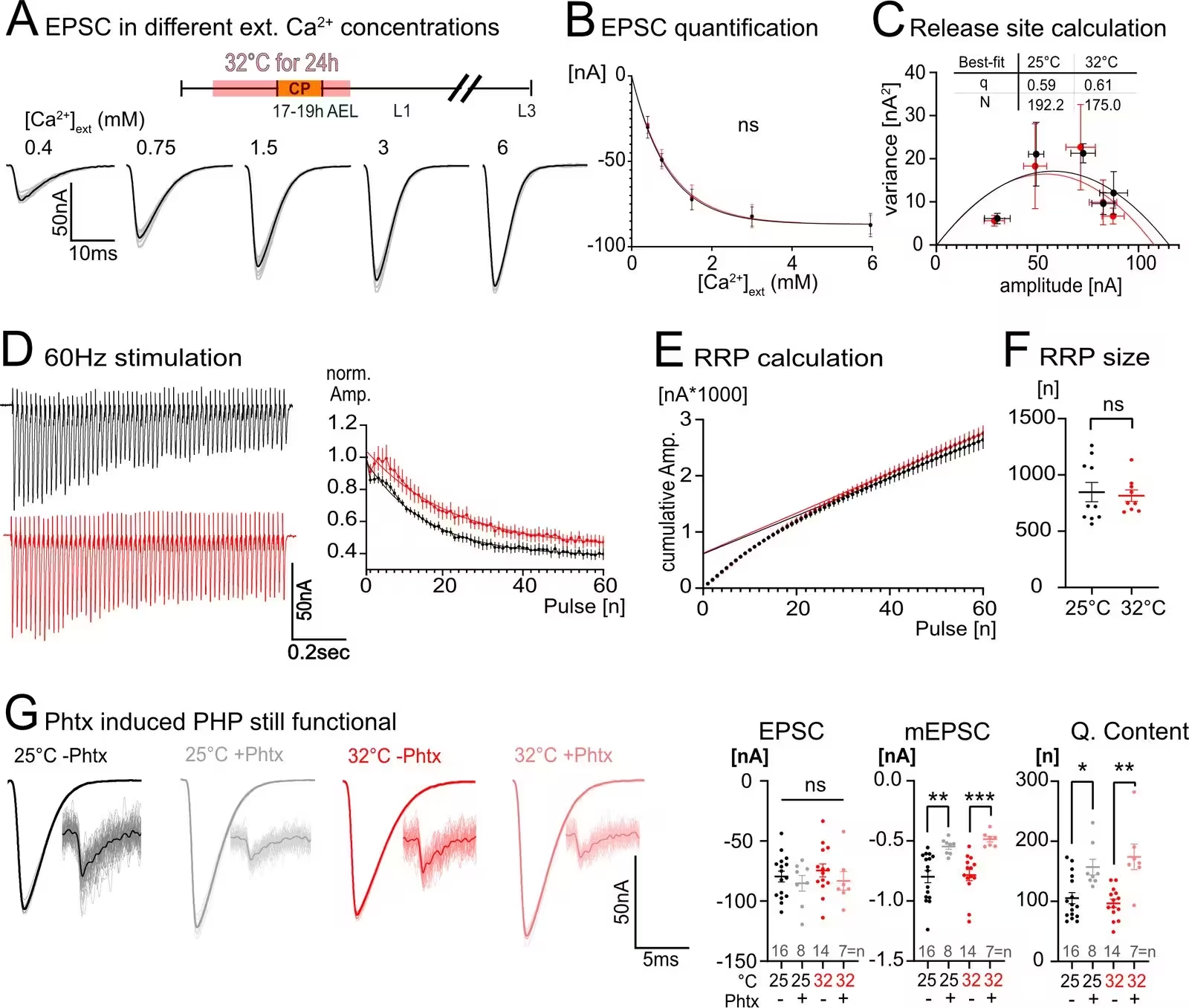
Figure 4: Physiological properties of the late larval neuromuscular junction (NMJ) following an embryonic 32 °C perturbation. (A) Representative traces of control two-electrode voltage clamp recordings, one per specimen, across a gradient of external calcium concentrations. (B) Postsynaptic current amplitude is not altered between control and embryonic heat stress-manipulated animals at any given external calcium concentration (n=5). (C) Plotting postsynaptic current amplitude against its variance to calculate quantal size (q) and the number of release sites being used (N), also did not show a significant difference between control and embryonic heat stress-manipulated animals (n=5). (D) To measure readily releasable pool size, a 60 Hz stimulus train was applied for 1 s where we could observe a slight increase in facilitation over the first 10 pulses in specimens that in their embryonic stage had experienced heat stress of 32 °C (n=9) as compared to controls (n=11). (E) Cumulative amplitude was
Physiological properties of the late larval neuromuscular junction (NMJ) following an embryonic 32 °C perturbation. (A) Representative traces of control two-electrode voltage clamp recordings, one per specimen, across a gradient of external calcium concentrations. (B) Postsynaptic current amplitude is not altered between control and embryonic heat stress-manipulated animals at any given external calcium concentration (n=5). (C) Plotting postsynaptic current amplitude against its variance to calculate quantal size (q) and the number of release sites being used (N), also did not show a significant difference between control and embryonic heat stress-manipulated animals (n=5). (D) To measure readily releasable pool size, a 60 Hz stimulus train was applied for 1 s where we could observe a slight increase in facilitation over the first 10 pulses in specimens that in their embryonic stage had experienced heat stress of 32 °C (n=9) as compared to controls (n=11). (E) Cumulative amplitude was Credit: Niklas Krick et al. / eLife

Sequential Maturation Windows Shape Organismal Behavior

The investigation further revealed that different tissue layers within the motor system possess their own distinct critical periods that open sequentially during embryogenesis. Developing body wall muscles exhibited a sensitivity to heat stress between 13 and 16 hours after egg laying, which selectively altered postsynaptic receptor composition without affecting larval movement. Only when heat stress struck the subsequent 17 to 19 hour window governing central locomotor circuitry did animals manifest behavioral deficits in crawling speed and seizure recovery.

These findings suggest that environmental stressors like high ambient temperatures leave lasting signatures on developing animals by altering the functional balance of central premotor networks. While peripheral junctions deploy robust homeostatic safeguards to maintain baseline transmission, central circuit alterations dictate broader behavioral outcomes, providing a clear cellular hierarchy for how neural networks adapt to ecological stress.

The research was published in eLife on October 26, 2026.

Fact Checked

This article has been fact checked for accuracy, with information verified against reputable sources. Learn more about us and our editorial process.

Last reviewed on .

Article history

  • Latest version

Reference(s)

  1. Krick, Niklas., et al. “Heterogeneous responses to embryonic critical period perturbations within the Drosophila larval locomotor circuit.” eLife, vol. 14, October 26, 2026 eLife Sciences Publications, Ltd, doi: 10.7554/eLife.108656. <https://doi.org/10.7554/eLife.108656>.

Cite this page:

Brown, Robert. “Embryonic Heat Waves Leave Permanent Scars on Developing Fly Brains.” BioScience. BioScience ISSN 2521-5760, 27 October 2026. <https://www.bioscience.com.pk/en/subject/biotechnology/heterogeneous-responses-to-embryonic-critical-period-perturbations-within-the-drosophila-larval-locomotor-circuit>. Brown, R. (2026, October 27). “Embryonic Heat Waves Leave Permanent Scars on Developing Fly Brains.” BioScience. ISSN 2521-5760. Retrieved October 27, 2026 from https://www.bioscience.com.pk/en/subject/biotechnology/heterogeneous-responses-to-embryonic-critical-period-perturbations-within-the-drosophila-larval-locomotor-circuit Brown, Robert. “Embryonic Heat Waves Leave Permanent Scars on Developing Fly Brains.” BioScience. ISSN 2521-5760. https://www.bioscience.com.pk/en/subject/biotechnology/heterogeneous-responses-to-embryonic-critical-period-perturbations-within-the-drosophila-larval-locomotor-circuit (accessed October 27, 2026).
End of the article