Enzyme Discovered That Stops Fatal Kidney Cell Death During Sepsis
Researchers reveal how a mitochondrial protein protects against sepsis-induced organ failure by regulating iron and fat storage.
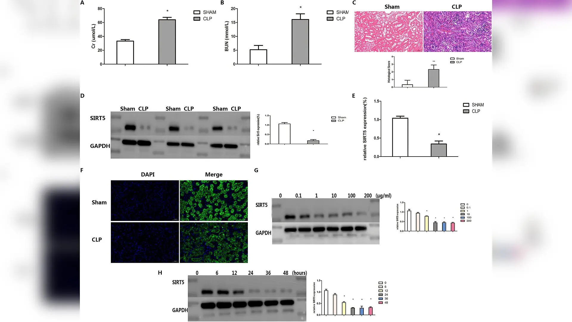
Sepsis triggers a severe form of kidney damage known as sepsis-associated acute kidney injury, which frequently leads to organ failure and high mortality rates in intensive care units. Scientists have struggled to develop targeted treatments because the exact molecular triggers inside the kidneys remain poorly understood. A new study reveals that a mitochondrial enzyme called SIRT5 acts as a crucial molecular shield against this type of kidney damage by stopping a destructive, iron-driven cell death process known as ferroptosis.
How Sepsis Destroys Kidney Protection
To understand the mechanisms behind this organ failure, the research team used both mouse models of sepsis and cultured human proximal tubular epithelial cells stimulated with lipopolysaccharide, which is a toxin that mimics bacterial infection. The study, published in PLOS One, demonstrated that levels of the protective enzyme SIRT5 plummet during sepsis. Normal kidneys showed abundant SIRT5 in the renal tubules, but septic mice and toxin-exposed cells exhibited a sharp, time- and dose-dependent drop in SIRT5 expression. This loss of SIRT5 crippled the kidney cells’ ability to handle oxidative stress.
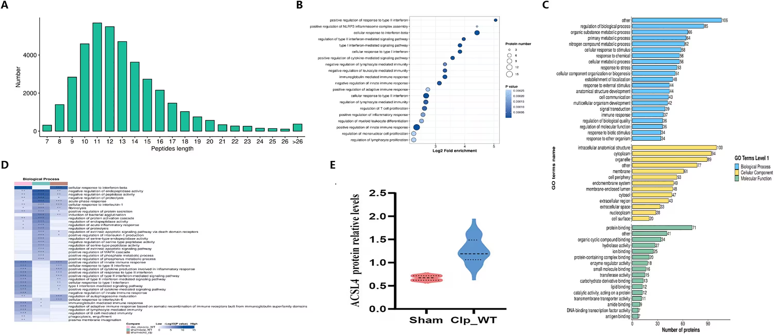
Ferroptosis is a specialized form of cell death caused by the toxic accumulation of iron and lipid peroxides within cell membranes. When the researchers performed quantitative proteomic analysis on kidney tissue, they discovered a massive disruption in iron homeostasis and ferroptosis pathways during sepsis. Specifically, septic conditions caused a severe increase in lipid reactive oxygen species, malondialdehyde, and ferrous iron, alongside a dangerous depletion of the protective antioxidant glutathione.
Restoring SIRT5 Reverses Kidney Damage
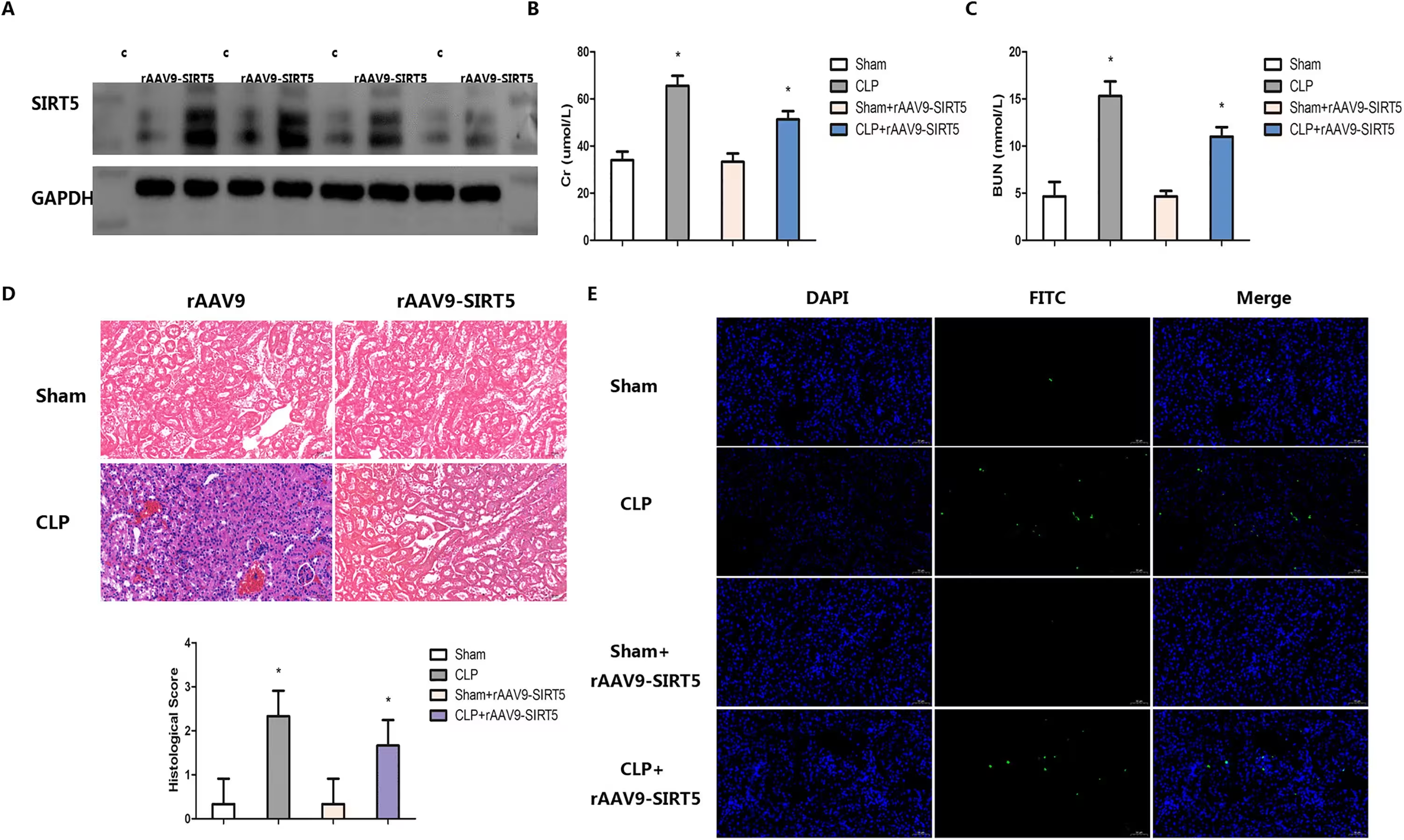
To test whether restoring this enzyme could halt the damage, the team used adeno-associated virus vectors to overexpress SIRT5 in mice before inducing sepsis. Elevating SIRT5 levels significantly preserved renal architecture, reduced blood markers of kidney injury such as creatinine and blood urea nitrogen, and lowered the number of apoptotic cells in the renal cortex. Conversely, mice engineered to lack SIRT5 specifically in their renal tubules suffered greatly accelerated kidney necrosis, higher injury scores, and massive accumulation of cellular debris.
Transmission electron microscopy revealed that sepsis caused kidney cell mitochondria to shrink, lose their internal membranes called cristae, and show increased membrane density. Overexpressing SIRT5 rescued these mitochondrial defects, whereas deleting SIRT5 made the structural collapse worse. Furthermore, treating injured cells with a specialized ferroptosis inhibitor called Ferrostatin-1 successfully blocked the excess cell death, proving that ferroptosis is the primary weapon of destruction in SIRT5-deficient kidneys.
The Molecular Axis Controlling Iron and Fats
The study mapped out the exact proteins involved in this protective pathway, pointing to a critical signaling axis composed of ACSL4 and FTH1. ACSL4 is an enzyme that actively promotes ferroptosis by packing cell membranes with polyunsaturated fatty acids that easily undergo harmful peroxidation. FTH1 is a major iron-storage protein that traps loose iron atoms and protects cells from iron-catalyzed oxidative destruction.
When SIRT5 was missing, ACSL4 protein levels spiked while FTH1 and antioxidant enzyme levels plummeted, creating a biochemical environment tailor-made for ferroptosis. When researchers knocked down ACSL4 in SIRT5-deficient cells, lipid peroxidation dropped, iron overload subsided, and cell viability returned. These experiments confirm that SIRT5 protects the kidneys by keeping the ACSL4 and FTH1 proteins in a healthy balance, preventing the fatal cascade of lipid and iron toxicity.
Limitations and Future Clinical Goals
While these findings highlight a promising pathway for treating septic kidney injury, several limitations must be noted. The research was conducted exclusively in mice and cultured cells, meaning that findings must still be validated in human clinical samples. Additionally, the transmission electron microscopy analysis was qualitative rather than quantitative, and the exact biochemical steps by which SIRT5 modifies the ACSL4 and FTH1 proteins require further investigation. Future therapies aiming to boost SIRT5 activity or block ACSL4 could eventually offer new ways to protect vulnerable patients from organ failure during severe infections.
The research was published in PLOS One on October 27, 2026.
This article has been fact checked for accuracy, with information verified against reputable sources. Learn more about us and our editorial process.
Last reviewed on .
Article history
- Latest version
Reference(s)
- Jiang, Liliang., et al. “SIRT5 deficiency exacerbates sepsis acute renal injury by activating ACSL4/FTH1 axis dependent ferroptosis.” PLOS One, vol. 21, no. 10, October 27, 2026, pp. e0361332 Public Library of Science (PLoS), doi: 10.1371/journal.pone.0361332. <https://doi.org/10.1371/journal.pone.0361332>.
Cite this page:
- Posted by Robert Brown