Advanced Computer Models Transform Targeted Cancer Drug Design
Sophisticated physiological simulations are helping researchers predict the complex behavior of antibody-drug conjugates while reducing reliance on animal testing.
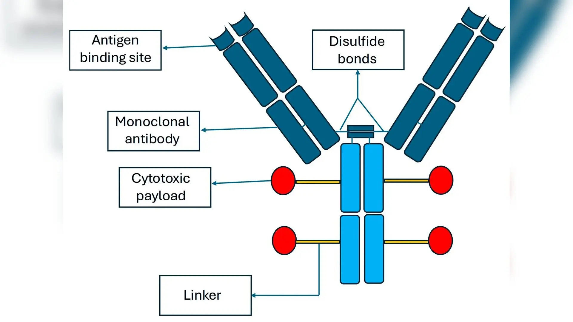
Complex biological cancer treatments called antibody-drug conjugates combine targeted antibodies with potent cytotoxic payloads, but predicting their behavior in the human body has long challenged researchers. A detailed review published in Toxics by Kanchan Bharti and colleagues outlines how sophisticated physiological computer simulations are transforming the development and safety assessment of these multi-analyte therapies.
Antibody-drug conjugates, commonly known as ADCs, function as guided missiles against cancer cells. They consist of a monoclonal antibody that zeroes in on specific antigens on tumor surfaces, a chemical linker, and a small-molecule drug payload that destroys the cell. While this design spares healthy tissue better than conventional chemotherapy, many candidates fail in clinical trials due to a narrow therapeutic index. This delicate balance means the dose required for a therapeutic response is uncomfortably close to the dose causing severe toxicity.
Why Traditional Pharmacokinetics Fall Short for ADCs
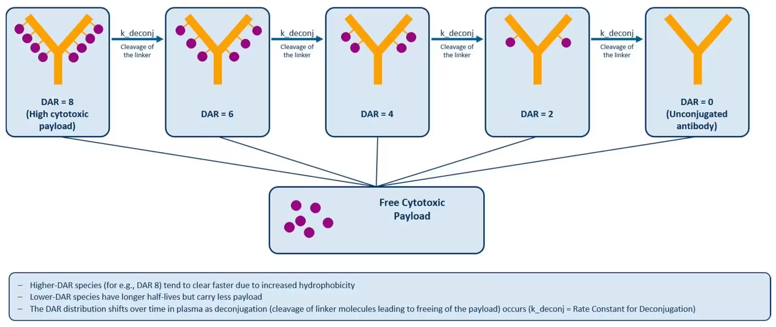
Conventional drug evaluation methods treat a medicine as a single, uniform substance moving through the body. However, an ADC is actually a complex mixture of intact conjugates, total antibodies, conjugated payloads, and free payloads all circulating simultaneously. Once administered, the chemical linkers can degrade prematurely in the bloodstream, releasing the toxic payload early and damaging healthy organs.
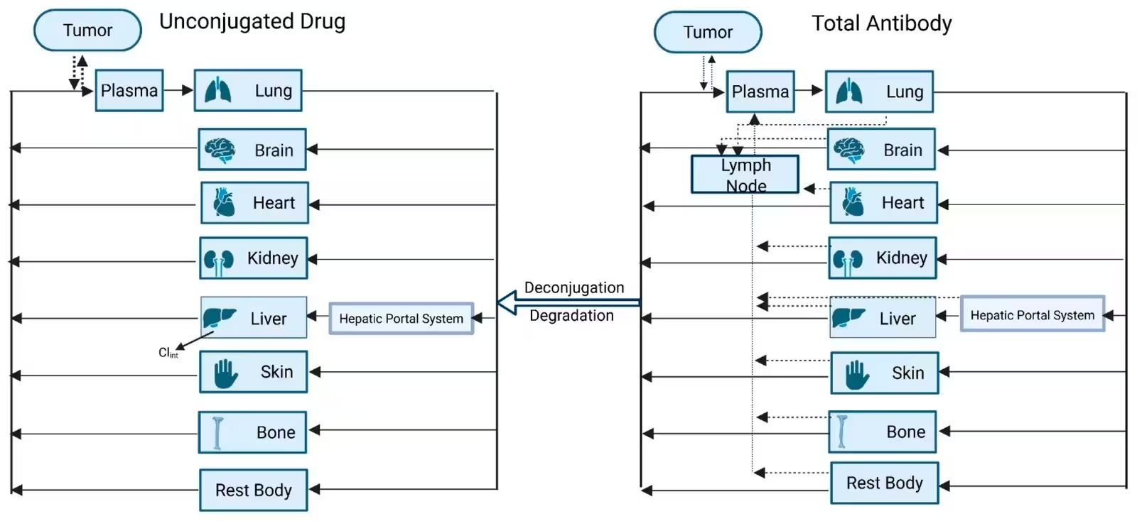
To capture this dynamic reality, pharmacologists rely on physiologically based pharmacokinetic modeling. This mathematical framework maps out how a drug distributes across different organs by simulating real biological processes. Unlike traditional methods, these advanced models divide tissue architecture into four distinct sub-compartments: vascular spaces, endothelial layers, interstitial fluid surrounding cells, and cellular interiors.
How Four-Layer Tissue Models Track Drug Movement
The vascular compartment handles initial delivery through blood plasma, where proteins create oncotic pressure that governs how fluids and large molecules filter out of capillaries. The endothelial layer acts as a physical barrier featuring different pore types depending on the organ, ranging from tight continuous capillaries in muscle tissue to wide open sinusoidal gaps in the liver. Within these endothelial cells, specialized receptors known as neonatal Fc receptors rescue antibodies from degradation and recycle them back into circulation, explaining why immunoglobulin G proteins enjoy a long plasma half-life of about 21 days.
Once large molecules reach the interstitial space between cells, they rely on convective fluid flow and pore transport. Cellular internalization then sorts the drug complexes, directing some back to the surface while routing others to lysosomes for payload release. Meanwhile, the released small-molecule payload behaves independently, crossing cell membranes to attack target tumors or diffusing into neighboring cells to trigger a bystander killing effect.
Integrating New Approach Methodologies and Artificial Intelligence
Regulatory bodies such as the United States Food and Drug Administration are actively encouraging the phasing out of animal testing in favor of alternative testing strategies. Physiologically based models fit directly into these new approaches by integrating laboratory cell assays and computer simulations to predict toxicity and drug-drug interactions before human trials begin. Researchers can forecast whether an ADC will experience dangerous metabolic interference when taken alongside other medications processed by liver enzymes.
Looking ahead, scientists are beginning to merge these mathematical models with artificial intelligence and machine learning to handle massive collections of molecular variables. By grouping similar structural features and running automated sensitivity analyses, machine learning tools can streamline complex calculations without sacrificing predictive accuracy. Although predicting precise drug concentrations deep inside human tumor tissue remains difficult due to limited clinical sampling, these computational frameworks offer a powerful path toward safer cancer treatments.
The research was published in Toxics on October 21, 2026.
This article has been fact checked for accuracy, with information verified against reputable sources. Learn more about us and our editorial process.
Last reviewed on .
Article history
- Latest version
Reference(s)
- Bharti, Kanchan., et al. “Mechanistic PBPK Modeling of Antibody–Drug Conjugates: Integration of Antibody Disposition, Linker Deconjugation, and Payload Pharmacokinetics.” Toxics, vol. 14, no. 10, October 21, 2026, pp. 931 MDPI AG, doi: 10.3390/toxics14100931. <https://doi.org/10.3390/toxics14100931>.
Cite this page:
- Posted by Robert Brown