Molecular Switch Driving Lymphatic Pumping Under Pressure Revealed
Researchers discover that lymphatic muscle cells rely on a specialized calcium signaling axis rather than traditional stretch channels to regulate fluid transport.
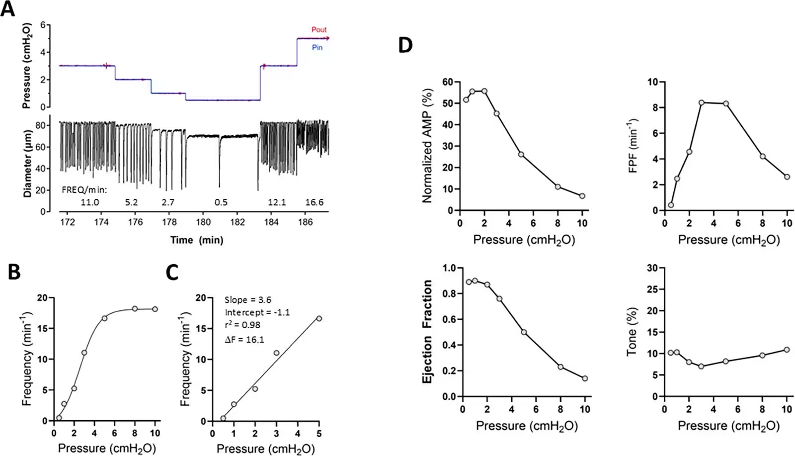
When fluid pressure rises inside tiny mouse lymphatic vessels, specialized muscle cells in their walls instantly speed up their pumping rhythm, jumping from 2 contractions per minute up to 20 contractions per minute over a narrow 5 centimeter of water pressure range. This vital homeostatic mechanism, known as pressure-induced lymphatic chronotropy, ensures that lymph fluid draining from tissues is efficiently propelled back into the bloodstream without building up in the body. Yet the precise molecular switches that translate physical stretch into electrical impulses have remained a major puzzle for physiologists studying the lymphatic system.
To uncover how these vessels sense pressure, a team of researchers led by scientists at the University of Missouri utilized advanced pressure myography, transgenic mouse models, and single-cell RNA sequencing to systematically test potential mechanosensory pathways. Their findings were published in eLife, revealing that lymphatic muscle cells rely on a specific signaling cascade rather than traditional stretch-activated ion channels found in other tissues.
Testing 16 Ion Channels Reveals Unexpected Culprits
Because lymphatic muscle cells share striking similarities with arterial smooth muscle, the research team initially suspected that classical mechanosensitive cation channels might drive the response. In arteries, proteins such as TRPC6, TRPM4, PKD2, TRPV2, and ENaC frequently mediate stretch-induced constriction. However, by examining mice with smooth muscle-specific knockouts for these various genes, the authors discovered that none of these channels are required for pressure-dependent lymphatic pacing.
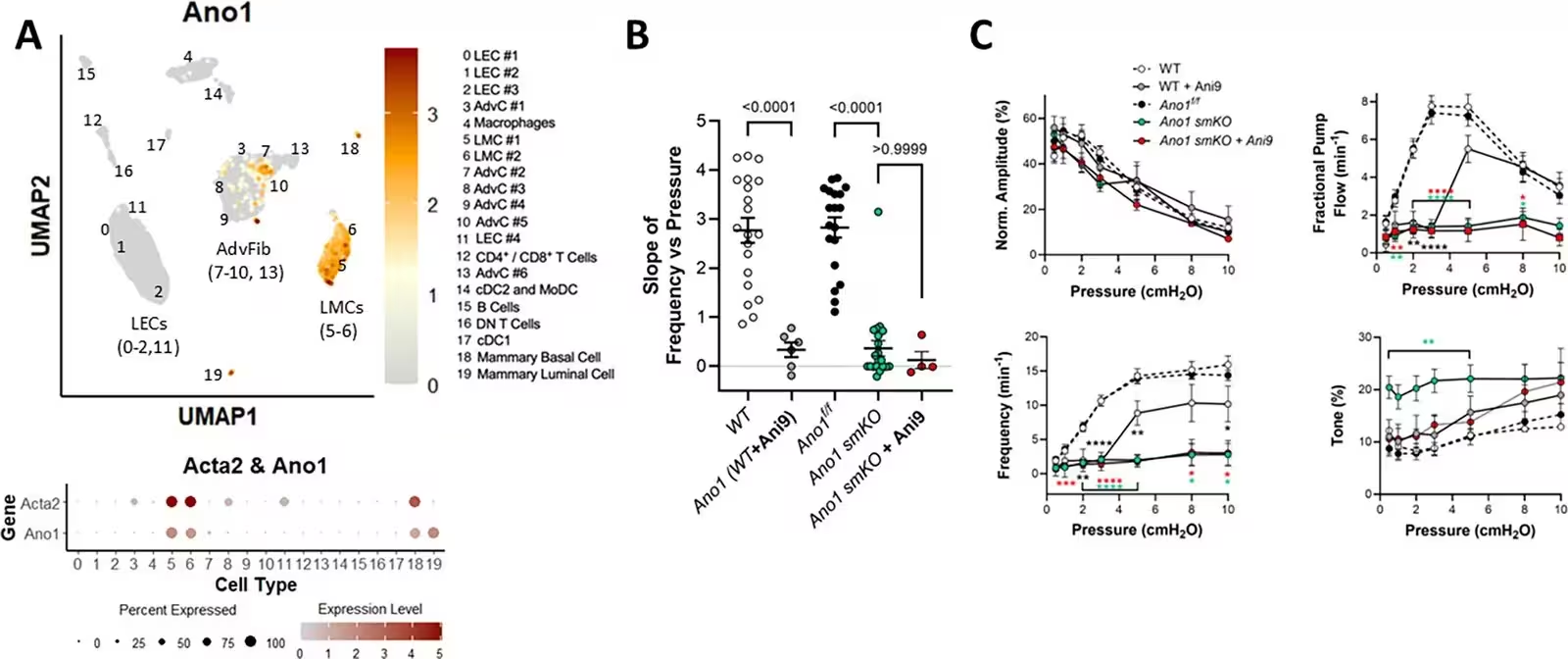
Even Piezo1, a widely celebrated cellular force sensor in the cardiovascular system, proved dispensable when deleted specifically from lymphatic muscle or endothelial cells. Out of 16 different ion channels tested via genetic deletion or pharmacological inhibition, only the calcium-activated chloride channel ANO1 (also called TMEM16A) and the voltage-gated calcium channel Cav1.2 proved essential for generating the dynamic frequency response.
Tracing the Signal Upstream to G-Protein Receptors
With ANO1 identified as the primary downstream effector driving the pacemaker current, the investigators searched upstream to find what triggers the channel. They discovered that deleting Itpr1, the gene encoding the intracellular calcium release channel IP3R1, dramatically blunted pressure-induced chronotropy in mouse popliteal collecting vessels, mimicking the effect of ANO1 loss.
This pointed directly toward G-protein-coupled receptors, specifically those linked to Gq and G11 proteins, which stimulate the production of inositol trisphosphate to release stored calcium from the sarcoplasmic reticulum. When the team used smooth muscle-specific knockouts of Gnaq combined with global knockouts of Gna11, pressure-induced chronotropy was severely impaired. Pharmacological inhibition using the selective Gq/11 blocker YM254890 similarly flattened the frequency response.
Remaining Questions in Vascular Mechanobiology
Although the study successfully maps out a core signaling axis involving Gq/11 proteins, IP3R1, and ANO1 channels, identifying the exact receptor protein that senses the physical stretch continues to be challenging. Single-cell RNA sequencing revealed that lymphatic muscle cells express over 130 different G-protein-coupled receptors. When the researchers tested antagonists for seven top candidates, including receptors for endothelin, neuropeptide Y, and thromboxane, none abolished the chronotropic response.
The authors note that multiple receptors working across different pressure ranges, or perhaps novel mechanosensitive adhesion receptors identified in their transcriptomic data, may act as the primary force transducers. Understanding these pathways offers crucial context for treating lymphatic dysfunction, such as lymphedema and fluid balance disorders linked to metabolic stress or genetic mutations.
The research was published in eLife on November 18, 2026.
This article has been fact checked for accuracy, with information verified against reputable sources. Learn more about us and our editorial process.
Last reviewed on .
Article history
- Latest version
Reference(s)
- Davis, Michael J., et al. “A Gαq/11-IP3R1-ANO1 pacemaking axis underlies pressure-induced lymphatic chronotropy.” eLife, vol. 14, November 18, 2026 eLife Sciences Publications, Ltd, doi: 10.7554/eLife.109466. <https://doi.org/10.7554/eLife.109466>.
Cite this page:
- Posted by Priya Sharma