New AI Model Reconstructs What You See Directly From Your Brain Activity
New AI technology can now reconstruct images from human brain scans with unprecedented detail, marking a major breakthrough in neuroimaging interpretation.
Researchers have developed a sophisticated artificial intelligence model capable of reconstructing images from human brain activity with unprecedented clarity. The system, known as the Brain Interaction Transformer (BIT), addresses persistent challenges in neuro-decoding by successfully capturing intricate details like color, spatial orientation, and composition that have historically eluded similar technologies.
The study, which has been submitted to the International Conference on Learning Representations, marks a significant leap from earlier attempts at visual reconstruction. While previous models often produced fuzzy approximations of an object’s general category, this new approach yields images that more accurately reflect the specific features of what a subject is observing.
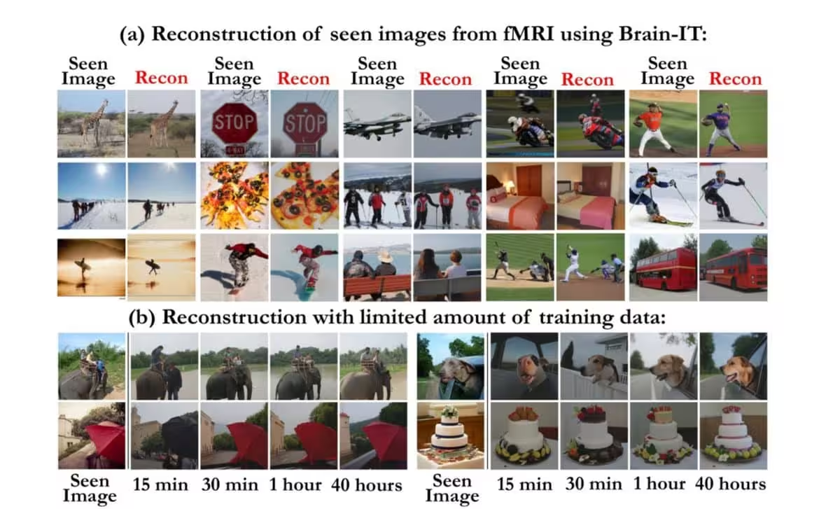
Overcoming the Data Bottleneck
A primary hurdle in training AI for fMRI analysis is the scarcity of paired data. Brain imaging sessions are time-consuming and expensive, resulting in limited datasets. The Weizmann Institute team utilized the Natural Scenes Dataset, which contains fMRI data from eight participants viewing roughly 73,000 images, but this volume remains insufficient for conventional deep learning requirements.
To bypass this constraint, the researchers engineered a bidirectional system. Brain-IT is capable of both decoding brain signals into visual outputs and predicting the specific neural responses an image would trigger. By creating a continuous feedback loop between random, unseen images and predicted fMRI signatures, the model effectively generates its own synthetic training data. This enables the AI to learn from a massive pool of visual information that was never part of the original human testing phase.
Mapping the Visual Cortex
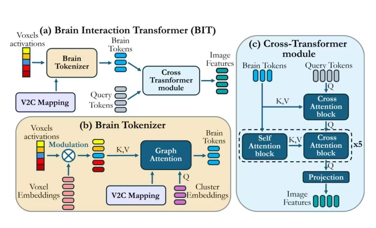
The Brain-IT architecture operates by tracking activity across approximately 40,000 voxels—the three-dimensional volumetric pixels used to map brain function. By isolating patterns of activation within these voxels, the model can differentiate between various visual stimuli, ranging from complex facial features to basic inanimate objects.
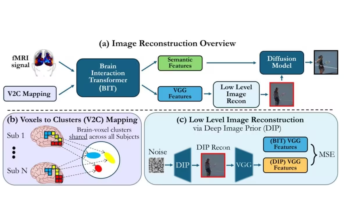
This granular approach has yielded unexpected insights into human neurobiology. During the development process, the system identified 128 distinct brain regions involved in image processing, some of which had not been previously categorized as central to visual perception in existing neurological literature.
Future Potential and Technical Frontiers
While the current iteration of Brain-IT is focused on static images, the researchers believe the technology holds significant promise for assistive communication, potentially aiding individuals living with severe physical paralysis. Furthermore, the ability to perturb images and observe subsequent fMRI changes offers a new, non-invasive method for neuroscientists to study how the brain reacts to specific visual variables.
Despite these successes, significant barriers remain. The temporal resolution of fMRI technology acts as a major bottleneck; the brain processes visual information in milliseconds, while a standard fMRI scan takes roughly two seconds to capture a snapshot. This disparity makes real-time video reconstruction an immense challenge. However, lead researcher Michal Irani suggests that if these temporal constraints can be overcome, the implications could be profound, potentially extending the technology to decode the imagery of human dreams.
This article has been fact checked for accuracy, with information verified against reputable sources. Learn more about us and our editorial process.
Last reviewed on .
Article history
- Latest version
Reference(s)
- Allen, Emily. “The natural scenes dataset: Massive, high-quality whole-brain 7T fMRI measurements during visual perception and memory.”, October 21, 2019 <https://naturalscenesdataset.org/>.
Cite this page:
- Posted by Asif Iqbal