New Artificial Intelligence Architecture Spots Hidden Colorectal Polyps With Greater Precision
A novel deep learning network combines spatial attention and frequency domain perception to help clinicians detect tricky intestinal lesions.
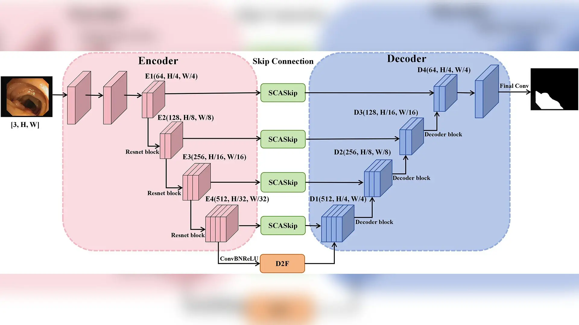
Catching tiny or folded colorectal polyps during a routine colonoscopy can be remarkably difficult for even the most experienced medical specialists, but a new artificial intelligence system is designed to spot these hidden growths with unprecedented precision. By combining advanced spatial attention with frequency domain analysis, the newly developed deep learning network addresses longstanding hurdles in medical imaging, such as uneven lighting, mucosal reflections, and blurry lesion edges.
How Cross-Scale Attention Refines Polyp Boundaries
Traditional computer vision models in colonoscopy analysis often struggle when merging feature information from different resolutions, leading to missed details and fuzzy boundaries. To overcome this, the researchers developed a Scale-Aware Cross Attention Skip Connection module, which acts as a bridge between the encoder and decoder stages of the network. Instead of treating all scales equally, this module uses multi-head attention and pyramid-based aggregation to synchronize features across multiple resolutions, allowing the model to capture polyps of vastly different physical sizes.
Furthermore, the architecture introduces a Dual-Domain Fusion module positioned at the bottleneck layer. This component separates low-frequency global context information from high-frequency fine details, such as texture and edges. By assigning adaptive weights to these separate frequency components, the model achieves a strong collaborative perception that makes vague and irregular polyp margins much more distinct.
Performance Benchmarks and Real-World Testing
To evaluate the effectiveness of the proposed architecture, the authors conducted extensive experiments using four publicly accessible benchmark repositories, including the CVC-ClinicDB and ETIS-LaribPolypDB collections. Detailed findings from these tests were published in PLOS One, demonstrating top-tier accuracy on standard evaluation metrics.
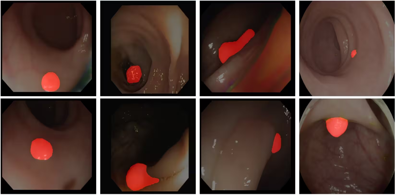
When tested on the CVC-ClinicDB dataset, the model achieved a mean Dice coefficient of 0.922 and a mean Intersection over Union of 0.863, outperforming several established segmentation frameworks. The system also maintained an inference speed of 5.93 milliseconds per image, yielding approximately 168 frames per second on high-performance hardware, which confirms its feasibility for real-time clinical assistance.
Current Limitations and Next Steps
Despite its high accuracy on familiar data distributions, cross-dataset evaluations revealed notable performance drops when the model encountered external datasets with substantial domain shifts, such as different imaging devices or complex illumination conditions. The authors noted that further work involving domain adaptation strategies and expanded data diversity will be necessary to ensure reliable clinical deployment across diverse hospital environments.
The research was published in PLOS One on November 20, 2026.
This article has been fact checked for accuracy, with information verified against reputable sources. Learn more about us and our editorial process.
Last reviewed on .
Article history
- Latest version
Reference(s)
- Gao, Rong., et al. “An enhanced U-shape architecture network with cross-scale attention and frequency-domain perception for robust colorectal polyp segmentation.” PLOS One, vol. 21, no. 11, November 20, 2026, pp. e0360491 Public Library of Science (PLoS), doi: 10.1371/journal.pone.0360491. <https://doi.org/10.1371/journal.pone.0360491>.
Cite this page:
- Posted by Asif Iqbal