MRI Study Finds Antisocial Traits Shrink Brain Volume by 1.5% in Psychopathic Men
Brain scans of 39 men with psychopathic traits reveal a distinct brain structure pattern, highlighting a unique trait dimension.

A team of German researchers investigated whether men with high psychopathic traits display measurable differences in brain anatomy, building on earlier behavioral assessments.
Data were drawn from three separate cohorts linked to the University Hospital Göttingen, Forschungszentrum Jülich, and the Medical Faculty of RWTH Aachen University. Because the scans originated from different sites, the researchers controlled for scanner type, acquisition protocol, age and intracranial volume in their statistical models.
Overall Brain Size Reduced in the Psychopathy Group
After adjusting for covariates, the psychopathic cohort showed a 1.45 % reduction in total brain volume compared with controls, with a nearly identical decrease on both hemispheres (1.50 % on the left, 1.40 % on the right). Broader reductions were also observed in the right telencephalon and cerebral cortex, surviving the study’s multiple‑testing correction.
Impulsive‑Antisocial Traits Linked to Widespread Volume Loss
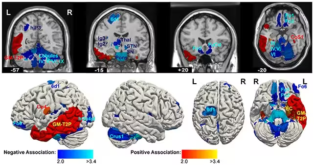
When the PCL‑R factors were examined separately, the dimension capturing impulsive and antisocial behaviour (Factor 2) displayed the most consistent structural pattern. Higher Factor 2 scores correlated with smaller volumes in a network of regions that includes the thalamus, subthalamic nucleus, brainstem nuclei, basal ganglia, orbitofrontal and dorsolateral prefrontal cortices, insular cortex and several cerebellar zones.
Even after correcting for the large number of statistical tests, the associations involving the subthalamic nucleus, left anterior thalamus, mesencephalon and pons remained statistically significant. The authors suggest that these findings may reflect alterations in frontal‑subcortical circuits implicated in behavioural regulation, though they caution that the results indicate correlation rather than causation.
In contrast, the interpersonal‑affective dimension (Factor 1) produced an inconsistent pattern. Some regions showed increased volume while others decreased, and none of these associations survived correction for multiple comparisons.
Before statistical correction, additional localized differences emerged in frontal, parietal and temporal cortices, the right insula, and parts of the hippocampal formation. A separate report highlighted the right subiculum as a notable area of change, but this specific finding did not retain significance after the full correction procedure.
Methodological Considerations and Future Directions
Structural MRI and deformation‑based morphometry were employed, primarily using the Julich‑Brain Atlas supplemented with regions from the AAL3 atlas. The heterogeneity of acquisition protocols across sites, the lack of uniform cognitive testing, and possible influences of substance use represent notable limitations.
The authors emphasize that the clearest neuroanatomical distinction aligns with the impulsive‑antisocial facet of psychopathy, and they call for further research to disentangle hereditary contributions from environmental effects.
This article has been fact checked for accuracy, with information verified against reputable sources. Learn more about us and our editorial process.
Last reviewed on .
Article history
- Latest version
Cite this page:
- Posted by Zubair Ali