Brain-Computer Interface Therapy Boosts Arm Recovery in Subacute Stroke Patients
A controlled trial shows that linking neural intent to robotic arm movement improves motor function and brain connectivity after stroke.
Thirty-two patients recovering from stroke who received active brain-computer interface therapy experienced significantly greater improvements in arm movement than those in a control group, a new randomized controlled trial indicates. The rehabilitation approach uses thought-controlled robotic assistance to help rebuild lost motor skills during the crucial months following a stroke.
Stroke frequently leaves survivors with persistent weakness in one arm, severely limiting their daily independence. The study focused on patients in the subacute phase, a critical window lasting one to three months post-stroke when the brain exhibits an enhanced capacity to reorganize and form new neural connections through experience-dependent learning.
How Thought-Controlled Robotics Assist Post-Stroke Arm Recovery
Researchers at Zhejiang Provincial People’s Hospital designed a participant-blinded trial to test whether connecting a patient’s movement intentions directly to robotic feedback accelerates recovery. In the active treatment group, patients wore an electroencephalography cap that recorded brain activity while they imagined performing swimming-like arm movements while watching a virtual game. When their neural signals crossed a specific threshold, a motorized robotic orthosis physically moved their arm.
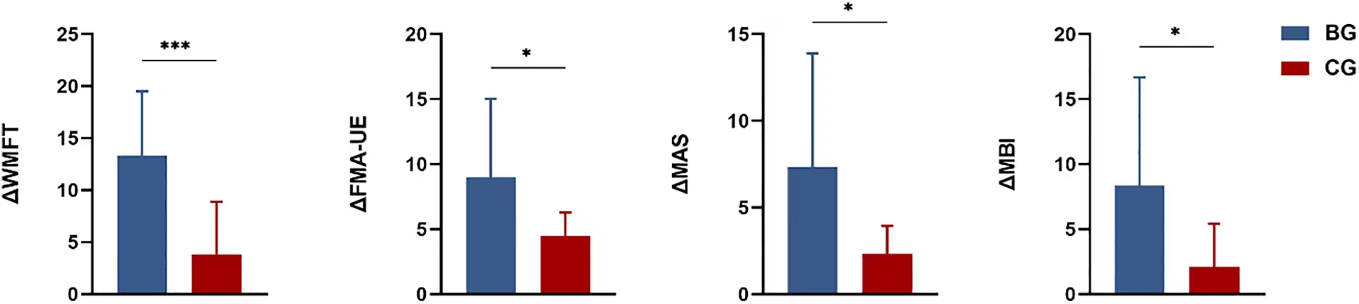
By contrast, the control group underwent an identical setup and training routine, but their brain signals were disconnected from the robotic hardware. Their arms were moved by preprogrammed robotic patterns without relying on their neural intent. Both groups completed 30-minute training sessions five days a week for four consecutive weeks alongside their standard physical and occupational therapy.
Measuring Clinical Motor Improvements and Brain Connectivity
To evaluate patient outcomes, the clinical team utilized the Fugl-Meyer Assessment-Upper Extremity scale alongside other standard motor and functional independence tests. The work was published in PLOS One by Qifeng Tong and colleagues, detailing how active brain-computer interface training produced superior score gains compared to the sham intervention.
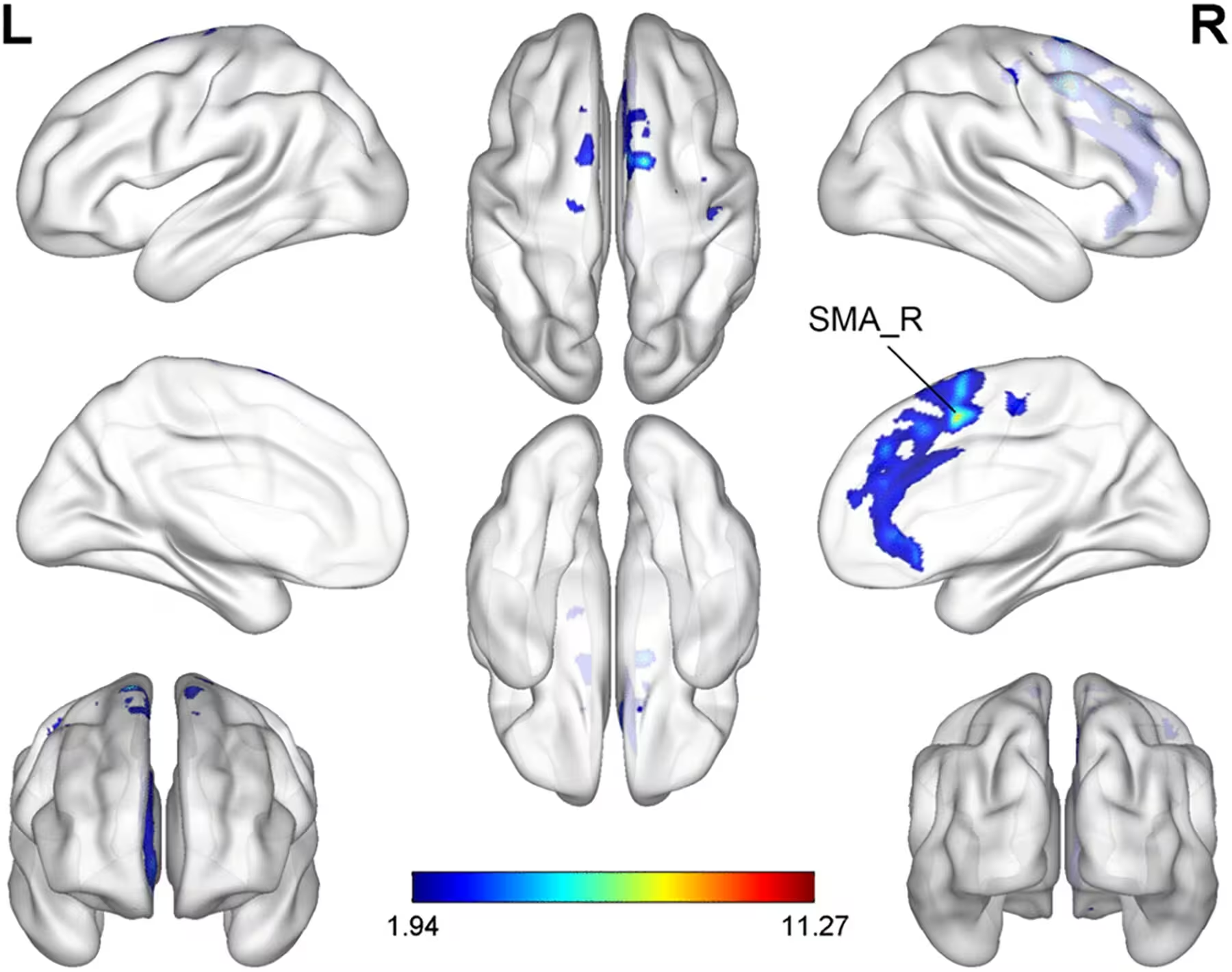
Alongside clinical testing, the researchers utilized structural and functional magnetic resonance imaging scans to track brain changes. The scans revealed that patients in the active training group developed stronger functional connectivity between a brain region called the right supplementary motor area, which plans movements, and other areas involved in sensory and motor processing, such as the cerebellum and fusiform gyrus. Furthermore, these enhanced neural connections correlated positively with the degree of physical recovery observed in the patients.
Study Limitations and What Lies Ahead for Brain-Computer Rehabilitation
Despite the promising results, the authors noted several important limitations. The trial involved a relatively small sample of 32 participants, which restricted statistical power and left some exploratory neuroimaging findings underpowered. Additionally, the intervention lasted only four weeks, and the researchers did not track long-term durability to see if the motor gains persisted months after the training stopped.
Future research with larger patient cohorts and extended follow-up periods will be necessary to confirm the long-term clinical benefits and help identify which specific patient subgroups benefit most from brain-computer interface rehabilitation.
The research was published in PLOS One on November 20, 2026.
This article has been fact checked for accuracy, with information verified against reputable sources. Learn more about us and our editorial process.
Last reviewed on .
Article history
- Latest version
Cite this page:
- Posted by Tariq Hassan