Sugar Modifications Fail to Stop Immune Reactions Against Brain-Targeting Drug Delivery Proteins
Medicine

Sugar Modifications Fail to Stop Immune Reactions Against Brain-Targeting Drug Delivery Proteins

Adding carbohydrate chains to antibody fragments failed to prevent anti-drug antibodies in mice, according to a new study.
By Sana Malik
Published:
Email this Article
Sugar Modifications Fail to Stop Immune Reactions Against Brain-Targeting Drug Delivery Proteins
Photo by Brecht Corbeel on Unsplash

Engineered antibody fragments designed to cross the blood-brain barrier trigger unwanted immune reactions in mice regardless of whether researchers add protective sugar molecules to their structure, according to a study evaluating protein therapeutics. Biopharmaceuticals represent a major class of modern treatments, but patients frequently develop anti-drug antibodies that neutralize the medication and cause complications.

How Sugar Modifications Failed to Block Immune Responses

To investigate whether adding specific carbohydrate chains could quiet this immune reaction, a team of researchers from Uppsala University and Karolinska Institutet focused on a model protein known as the single-chain variable fragment of the 8D3 antibody, or scFv8D3. This specialized fragment helps large therapeutic molecules enter the brain by latching onto transferrin receptors on the cellular lining of blood vessels. When researchers modified the protein to carry extra N-linked glycosylation sites packed with terminal sialic acid residues, the modified fragments maintained their ability to bind to the transferrin receptor. The work is published in PLOS One.

Terminal sialic acid groups are known to engage regulatory receptors on immune cells and can sometimes suppress unwanted inflammation or extend the circulation time of therapeutic proteins in the bloodstream. Because natural antibodies sometimes carry these sugar structures in their antigen-binding regions, the researchers hypothesized that engineering similar sites onto the scFv8D3 fragment would mask vulnerable protein surfaces and suppress the production of neutralizing antibodies.
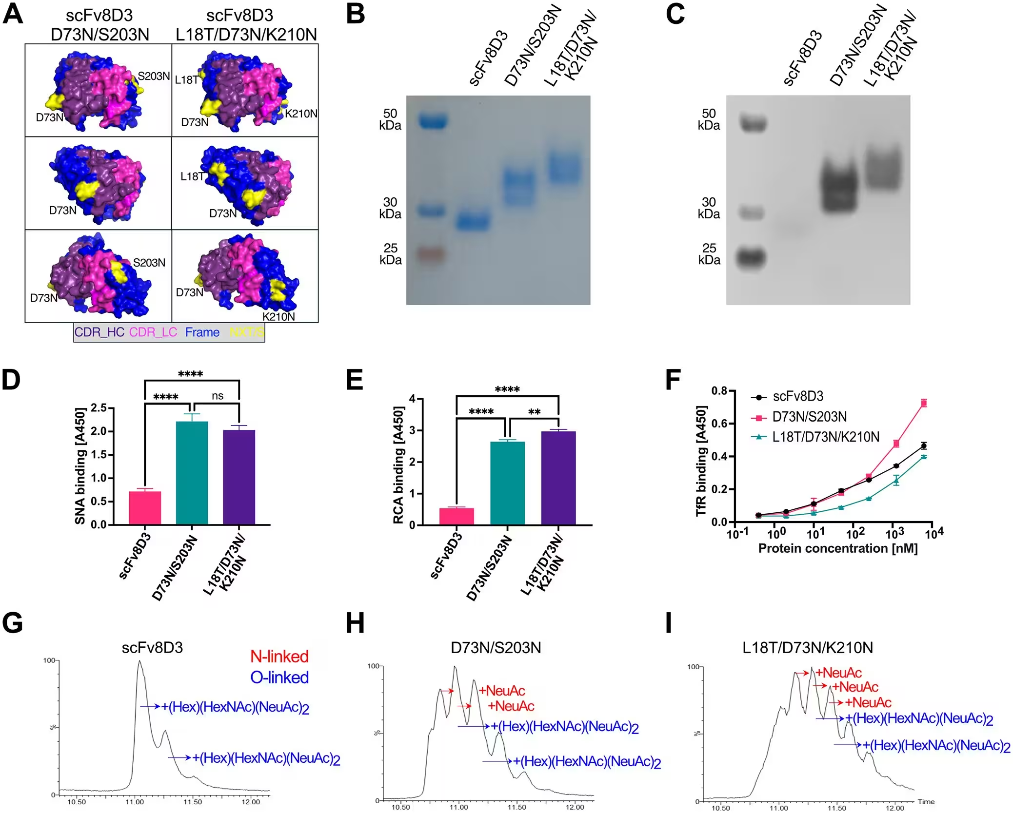
Fig 1: Design and characterization of glycosylated scFv8D3 variants. (A) AlphaFold 3 structure of scFv8D3 visualized in PyMOL, with complementarity-determining regions (CDR), framework regions, and positions of the inserted glycosylation motifs (NXT/S) highlighted. (B) SDS-PAGE analysis of Strep-tag purified scFv8D3 and glycosylated variants under non-reducing conditions. A pre-stained protein ladder was used to approximate molecular weights, which are predicted to be 29.6 kDa for all three variants based on the amino acid sequence. (C) Lectin blot with Sambucus nigra agglutinin visualizing the presence of sialic acid residues in the scFv8D3 variants. (D) Sambucus nigra agglutinin (SNA) binding ELISA demonstrating higher sialic acid presence in the N-linked glycosylation mutants. (E) Ricinus communis agglutinin (RCA) binding to N-linked galactose showing increased galactose levels in the mutants. (F) Indirect ELISA demonstrating binding of the protein variants to the transferrin receptor (TfR
Design and characterization of glycosylated scFv8D3 variants. (A) AlphaFold 3 structure of scFv8D3 visualized in PyMOL, with complementarity-determining regions (CDR), framework regions, and positions of the inserted glycosylation motifs (NXT/S) highlighted. (B) SDS-PAGE analysis of Strep-tag purified scFv8D3 and glycosylated variants under non-reducing conditions. A pre-stained protein ladder was used to approximate molecular weights, which are predicted to be 29.6 kDa for all three variants based on the amino acid sequence. (C) Lectin blot with Sambucus nigra agglutinin visualizing the presence of sialic acid residues in the scFv8D3 variants. (D) Sambucus nigra agglutinin (SNA) binding ELISA demonstrating higher sialic acid presence in the N-linked glycosylation mutants. (E) Ricinus communis agglutinin (RCA) binding to N-linked galactose showing increased galactose levels in the mutants. (F) Indirect ELISA demonstrating binding of the protein variants to the transferrin receptor (TfR Credit: Gustaf Hederoth et al. / PLOS One

What Happened During Animal Testing

The investigators tested both double-glycosylated and triple-glycosylated variants of the scFv8D3 protein in C57BL/6 mice. After confirming that the purified proteins successfully retained their structural stability and carried high levels of sialic acid and galactose, the team evaluated their distribution in the body. When administered as a single dose, the glycoengineered variants showed brain uptake comparable to the unmodified wild-type protein, though the modified constructs accumulated more readily in the liver.

When the researchers administered repeated doses over an extended period to induce a robust immune reaction, the outcome changed significantly. All treatment groups developed anti-drug antibodies, regardless of whether the protein variants carried the extra sugar modifications. Animals that mounted high immune responses cleared the circulating protein much faster than animals with low antibody titers, which severely compromised the brain-targeting capability of the drug delivery vehicle.
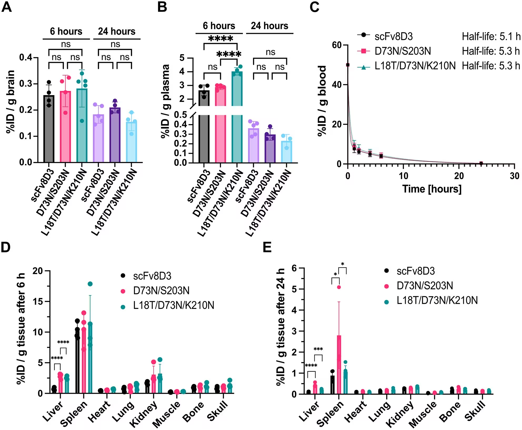
Fig 2: Effect of glycosylation on scFv8D3 pharmacokinetics and biodistribution. Brain uptake (A) and plasma levels (B) of radiolabelled scFv8D3 variants at 6- and 24-hours post-injection. (C) Blood pharmacokinetics of the scFv8D3 variants, determined from sequential blood samples and fitted using a two-phase decay non-linear regression model. (D,E) Biodistribution of the scFv8D3 variants 6 hours (D) and 24 hours (E) post-injection. Results are presented as mean±SD (n = 4-5 animals per group). Tissue radioactivity is expressed as percent injected dose per gram of tissue (%ID/g). Statistical significance was assessed using one-way ANOVA followed by Tukey’s post hoc test for pairwise comparisons. P-values were defined as follows: ns > 0.05; * < 0.05; ** < 0.01; *** < 0.001; **** < 0.0001.
Effect of glycosylation on scFv8D3 pharmacokinetics and biodistribution. Brain uptake (A) and plasma levels (B) of radiolabelled scFv8D3 variants at 6- and 24-hours post-injection. (C) Blood pharmacokinetics of the scFv8D3 variants, determined from sequential blood samples and fitted using a two-phase decay non-linear regression model. (D,E) Biodistribution of the scFv8D3 variants 6 hours (D) and 24 hours (E) post-injection. Results are presented as mean±SD (n = 4-5 animals per group). Tissue radioactivity is expressed as percent injected dose per gram of tissue (%ID/g). Statistical significance was assessed using one-way ANOVA followed by Tukey’s post hoc test for pairwise comparisons. P-values were defined as follows: ns > 0.05; * < 0.05; ** < 0.01; *** < 0.001; **** < 0.0001. Credit: Gustaf Hederoth et al. / PLOS One

Understanding the Limitations of Glycoengineering

While previous scientific literature has linked sialylation to the anti-inflammatory properties of pooled human immunoglobulins used in clinical treatments, the current findings indicate that these protective signals do not automatically prevent the formation of anti-drug antibodies against engineered antibody fragments. The simultaneous increase in terminal galactose residues on the modified proteins may have promoted clearance through hepatic receptors, counteracting any potential circulation benefits.

The study authors note that while their conclusions stem from the specific scFv8D3 model, the experimental design reflects the challenges of long-term biologic administration in clinical settings. Future investigations involving different therapeutic formats will help clarify whether carbohydrate engineering can be optimized for other classes of brain-targeting protein therapeutics.

The research was published in PLOS One on November 20, 2026.

Fact Checked

This article has been fact checked for accuracy, with information verified against reputable sources. Learn more about us and our editorial process.

Last reviewed on .

Article history

  • Latest version

Cite this page:

Malik, Sana. “Sugar Modifications Fail to Stop Immune Reactions Against Brain-Targeting Drug Delivery Proteins.” BioScience. BioScience ISSN 2521-5760, 20 November 2026. <https://www.bioscience.com.pk/en/subject/medicine/engineered-fab-glycosylation-of-a-blood-brain-barrier-transporter-single-chain-variable-fragment-preserved-functionality-but-did-not-impact-anti-drug-antibody-formation>. Malik, S. (2026, November 20). “Sugar Modifications Fail to Stop Immune Reactions Against Brain-Targeting Drug Delivery Proteins.” BioScience. ISSN 2521-5760. Retrieved November 20, 2026 from https://www.bioscience.com.pk/en/subject/medicine/engineered-fab-glycosylation-of-a-blood-brain-barrier-transporter-single-chain-variable-fragment-preserved-functionality-but-did-not-impact-anti-drug-antibody-formation Malik, Sana. “Sugar Modifications Fail to Stop Immune Reactions Against Brain-Targeting Drug Delivery Proteins.” BioScience. ISSN 2521-5760. https://www.bioscience.com.pk/en/subject/medicine/engineered-fab-glycosylation-of-a-blood-brain-barrier-transporter-single-chain-variable-fragment-preserved-functionality-but-did-not-impact-anti-drug-antibody-formation (accessed November 20, 2026).
End of the article