Arc Protein Hijacked to Spread Toxic Tau: New Pathway Offers Fresh Alzheimer’s Target
Health

Arc Protein Hijacked to Spread Toxic Tau: New Pathway Offers Fresh Alzheimer’s Target

Study finds a brain protein may help Alzheimer’s spread via tiny vesicles, offering a fresh target to slow disease progression.

By David Anderson
Published:
Email this Article
Brain Protein Discovery Reveals Hidden Pathway That May Drive Alzheimers Spread Scaled
Credit: Shutterstock | Dungrela Publishing

New research published in Cell points to a previously unknown cellular route that may change how scientists view the spread of Alzheimer’s disease across the brain. The study identifies a normal brain protein that appears to unintentionally ferry toxic Tau, the protein closely linked to neuronal loss and memory decline, between cells. The findings imply that disease progression depends not only on the accumulation of harmful proteins inside neurons but also on the mechanisms that move these proteins from one cell to another.

By facilitating intercellular transfer, this pathway could speed the propagation of pathology throughout different brain regions over time. Experiments in laboratory models showed that the protein Arc plays a pivotal role in packaging and shuttling Tau from one neuron to the next, suggesting that a communication system essential for healthy brain function may be co‑opted in disease.

Arc Bridges Normal Signalling and Disease Transmission

The investigation, detailed in Cell, highlights Arc as a double‑edged molecule that supports both regular neuronal activity and the spread of Alzheimer‑related damage. Under normal circumstances, Arc helps neurons exchange signals via tiny membrane‑bound packets called extracellular vesicles, which travel across synapses to maintain brain homeostasis.

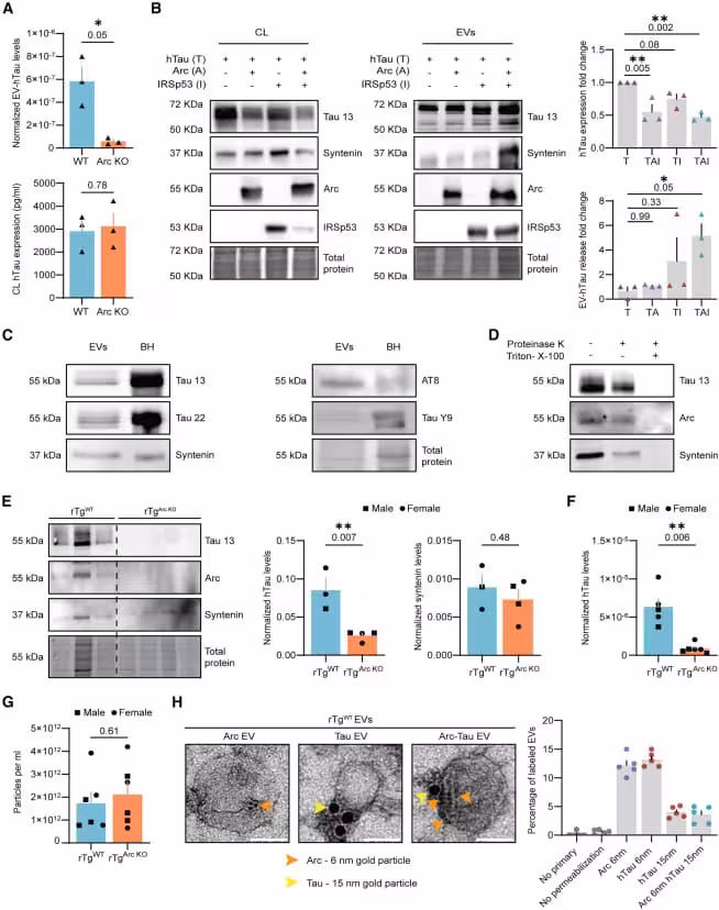
In Alzheimer’s models, the researchers found that toxic Tau hijacks this vesicle system. Rather than staying confined to the originating cell, Tau binds to Arc‑laden vesicles and is delivered to neighboring healthy neurons, where it can trigger further protein misfolding and cellular impairment. The data suggest that a network designed for communication can become a conduit for disease spread.

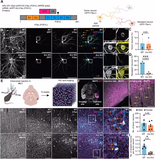
Comparisons between normal mice and those lacking Arc revealed a dramatic drop in Tau movement when Arc was absent, underscoring its central role in intercellular transmission.

(A) Arc facilitates the release of hTau in rat neuronal EVs. WT and Arc KO cortical primary cultures were transduced with hSyn-eGFP-2A-2N4R-hTau(P301L) lentivirus. Conditioned media and cell lysates (CL) were collected, and EVs were isolated using SEC. hTau levels in isolated EVs and CL were assessed using an ELISA. EV-hTau levels normalized to total protein (DC protein assay) were significantly reduced in Arc KO cultures, while hTau expression in CL was comparable between WT and Arc KO neurons (n = 3 independent experiments).
(B) hTau release is enhanced by Arc and IRSp53 overexpression in N2a cells. Neuro 2a cells were transfected with 2N4R-hTau(P301L) and Arc or IRSp53 or both. Cell lysates and conditioned media were collected 24 h after transfection. EVs were isolated using SEC. hTau release in EVs is enhanced when both Arc and IRSp53 are expressed, despite slightly lower levels of hTau in the cell lysate (n = 3 independent experiments).
(C) hTau is released in brain EVs isolated from rTgWT mice. EVs (F1-4 fractions were pooled after SEC from 3 male mice) and brain homogenates (BHs) from 4-month-old rTgWT mice were immunoblotted for hTau (Tau 13, Tau 22, AT8, and Tau Y9 antibodies) and syntenin. See also Figure S1 for brain EV characterization.
(D) hTau and Arc in EVs are protected from Proteinase K degradation. EVs were isolated from the brains of 4-month-old rTgWT mice (F1-4 fractions were pooled after SEC from 3 female mice) and incubated with Proteinase K (7 μg/mL) with or without detergent (1% triton-X-100) for 10 min. A representative western blot shows Arc, hTau, and syntenin are protected from Proteinase K degradation when no detergent is present.
(E and F) Arc facilitates the release of hTau in mouse brain EVs. EVs were isolated from the brains of 4-month-old rTgWT mice (n = 3 preps, 2M, 4F, with each EV prep consisting of two brains) and rTgArc KO (n = 4 preps, 4M, 4F). Pooled EV fractions were blotted for Tau 13, Arc, and syntenin. Dotted line indicates a spliced blot to crop out irrelevant lanes. Levels of hTau normalized to total protein were significantly reduced in the absence of Arc, while there was no difference in syntenin levels normalized to total protein. (F) hTau levels in EVs from 4-month-old rTgWT (n = 5 preps, 4M, 6F) and rTgArcKO (n = 6 preps, 6M, 6F) mice were normalized to total protein (DC protein assay) and quantified using total hTau ELISA. rTgArcKO EVs contain less hTau than rTgWT EVs.
(G) Total EV production is unaffected in rTgArc KO mice. EVs were isolated from the brains of 4-month-old rTgWT mice and rTgArc KO (n = 6 preps, 3M, 3F). Fractions 1–4 were pooled and analyzed by nanoparticle tracking analysis. There were no significant differences in the total number of EVs. See also Figure S2D.
(H) hTau and Arc are co-packaged in EVs isolated from rTgWT mice. EVs were isolated from 4-month-old rTgWT mice (n = 1 EV prep, 2F). The isolated EVs were either single immunogold labeled for Arc or hTau using 6 nm gold particles or double immunogold labeled using 6 nm gold particles for Arc and 15 nm gold particles for hTau. Representative TEM images of Arc, hTau, and Arc-hTau EVs. Scale bar, 50 nm. Quantification of the percentage of labeled EVs from 5 images is shown. See also Figure S2E for representative images. Data are represented as mean ± SEM. ∗p ≤ 0.05, ∗∗p ≤ 0.01 using a one-way ANOVA (B) or an unpaired t test (A and E–G).
Arc is critical for the release of hTau in neuronal EVs

How Toxic Tau Forms “Glue‑Like” Aggregates Inside Neurons

Once initiated, the process can cascade through neural networks, spreading damage in a ripple effect. The study demonstrates that Arc‑associated vesicles may act as carriers for these seeding fragments. Inside the vesicles, Tau is shielded from enzymatic breakdown, increasing the likelihood that it reaches new neurons. This mechanism offers a cellular explanation for the progressive loss of memory and cognition observed in Alzheimer’s patients.

 (A and B) EV-hTau seeds tau aggregation. HEK biosensor cells were transfected with 10 μg of purified recombinant hTau, as a positive control for FRET induction, or equal amounts (10 μg total protein) of rTgWT or rTgArcKO EVs isolated from 4-month-old mice. Cells transfected with purified hTau or rTgWT EVs show FRET-positive cells. Scale bar, 50 μm (main images), 10 μm (insets). (B) Equal amounts (10 μg total) of rTgWT (n = 3 preps, 2M, 4F) and rTgArcKO (n = 4 preps, 4M, 4F) EVs were transfected in HEK biosensor cells, and FRET-positive cells were counted using flow cytometry. FRET-positive HEK biosensor cells exhibiting hTau aggregation are observed in the FRET gate in the representative flow cytometry plots. There are fewer FRET-positive cells that were transfected with rTgArc KO EVs as compared with FRET-positive cells transfected with rTgWT EVs. The integrated FRET intensity (calculated by multiplying the percentage of FRET-positive cells and median fluorescence intensity of FRET-positive cells) of cells transfected with rTgArc KO EVs is significantly lower than cells transfected with rTgWT EVs, indicating a lack of seeding. Data are represented as mean ± SEM. ∗p ≤ 0.05 using an unpaired t test (B).
 Brain-derived EV-hTau seeds hTau aggregation

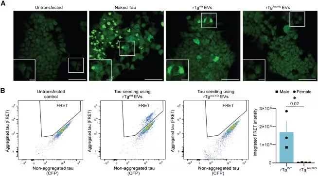
Mouse Experiments Show Sharp Decline in Tau Spread Without Arc

Data from mouse models support the proposed mechanism. Researchers compared brains that express normal levels of Arc with those where the protein was genetically removed. In the Arc‑deficient mice, the transfer of Tau between neurons fell dramatically, altering the trajectory of disease development. One of the investigators summed up the observation: “When we removed Arc, we saw that the transfer of Tau was severely, severely reduced. It was almost gone.” These results indicate that Arc may be required for efficient intercellular movement of Tau.

In the absence of Arc, toxic proteins tend to remain trapped inside affected neurons, potentially leading to higher intracellular toxicity while limiting spread to neighboring cells. This trade‑off highlights the challenge of designing therapies that block disease propagation without harming the neurons that rely on Arc for normal waste management.

Balancing Neuronal Protection and Pathology Propagation

Further analysis revealed a nuanced role for Arc in neuronal health. When active, Arc enables neurons to export excess Tau, reducing internal stress and extending cell survival. At the same time, the exported material can be taken up by nearby healthy cells, extending the pathological cascade.

These findings suggest that directly inhibiting Arc could worsen damage in already compromised cells. Consequently, researchers are exploring strategies that intercept vesicles after they leave the donor neuron rather than preventing their formation altogether.

Potential Impact on Future Alzheimer’s Therapies

The team also detected Arc‑ and Tau‑laden vesicles in human brain samples, hinting that the mechanism may operate beyond animal models. Nevertheless, the authors caution that many questions remain about how this process functions in people. “Most of the work we’ve been doing is in mice, not in humans,” Shepherd notes. “We have some clues that whatever is happening in these mice could also be happening in humans, but we don’t know that yet. And we’re far away from saying that we’re developing a treatment for anything.”

(A) Schematic of the intercellular hTau transmission assay. hSyn.eGFP-2A-2N4R-hTau(P301L) produces equal amounts of eGFP and hTau in donor neurons.
(B) Representative images of hTau and eGFP staining in WT and Arc KO hippocampal primary neurons. Neurons were sparsely transduced at DIV 7. 14 days post transduction, the neurons were fixed and immunostained for eGFP and hTau. Transduced donor neurons are indicated by yellow arrows, and recipient neurons are indicated by orange arrows. Scale bar, 100 μm (main images), 50 μm (zoomed in images).
(C) Virus transduction is similar in WT and Arc KO primary neurons. The percentage of donor neurons is not different in WT and Arc KO neurons, indicating comparable viral transduction (n = 3 independent cultures). See also Figure S10 for transduction at DIV7.
(D) Intercellular hTau transmission is reduced in Arc KO primary neurons. The percentage of recipient neurons is significantly reduced in Arc KO neurons (n = 3 independent cultures).
(E) Schematic for the in vivo tau transmission assay. For in vivo studies, 6-month-old WT and Arc KO mice were injected with the hSyn-eGFP-2A-2N4R hTau(P301L) virus unilaterally in the medial entorhinal cortex. 10 weeks post-injection, the brains were collected, sectioned, and immunostained for eGFP and hTau. Imaging was performed in the contralateral media entorhinal cortex.
(F) Representative images showing injection and imaging sites. Yellow arrows indicate donor neurons at the imaging site. Sparse transduction in the contralateral entorhinal cortex allows hTau transmission to be quantified. Scale bar, 100 μm.
(G) Representative images of hTau and eGFP staining in WT and Arc KO entorhinal cortex. Transduced donor neurons are indicated by yellow arrows, and recipient neurons are indicated by orange arrows. Scale bar, 100 μm (main images), 50 μm (zoomed in images).
(H) Virus transduction is similar in the WT and Arc KO mice’s entorhinal cortex. WT and Arc KO mice show no differences in the number of donor neurons, indicating comparable levels of virus transduction in WT and Arc KO mice (n = 6, 3M, 3F).
(I) Intercellular hTau transmission is reduced in Arc KO mice in vivo. The number of recipient neurons per donor neuron is reduced in Arc KO mice, indicating reduced intercellular hTau transmission (n = 6, 3M, 3F). Data are represented as the mean ± SEM. ∗p ≤ 0.05, ∗∗∗p ≤ 0.001 using an unpaired t test (C, D, H, and I). See also Figure S11 for electrophysiological analysis of Arc KO neurons.
 Arc is critical for intercellular hTau transmission

Shepherd adds that targeting the vesicles after they exit the donor cell could provide a therapeutic window: “If we could target these particular EVs, that would be a really useful therapy strategy. For someone with early‑onset Alzheimer’s or dementia, if we could stop the spread, then we could prevent further damage and cognitive decline.” This approach shifts the focus from removing toxic proteins to blocking their movement, opening a new avenue in Alzheimer’s research.

Fact Checked

This article has been fact checked for accuracy, with information verified against reputable sources. Learn more about us and our editorial process.

Last reviewed on .

Article history

  • Latest version

Cite this page:

Anderson, David. “Arc Protein Hijacked to Spread Toxic Tau: New Pathway Offers Fresh Alzheimer’s Target.” BioScience. BioScience ISSN 2521-5760, 30 June 2026. <https://www.bioscience.com.pk/en/subject/health/brain-protein-discovery-reveals-hidden-pathway-that-may-drive-alzheimers-spread>. Anderson, D. (2026, June 30). “Arc Protein Hijacked to Spread Toxic Tau: New Pathway Offers Fresh Alzheimer’s Target.” BioScience. ISSN 2521-5760. Retrieved June 30, 2026 from https://www.bioscience.com.pk/en/subject/health/brain-protein-discovery-reveals-hidden-pathway-that-may-drive-alzheimers-spread Anderson, David. “Arc Protein Hijacked to Spread Toxic Tau: New Pathway Offers Fresh Alzheimer’s Target.” BioScience. ISSN 2521-5760. https://www.bioscience.com.pk/en/subject/health/brain-protein-discovery-reveals-hidden-pathway-that-may-drive-alzheimers-spread (accessed June 30, 2026).

Follow us on social media

End of the article