Four Children With Rare Brain Iron Disorders Reveal Hidden Diagnostic Hurdles
Genetic testing and advanced scans uncovered pantothenate kinase-associated and mitochondrial membrane protein-associated neurodegeneration in young patients.
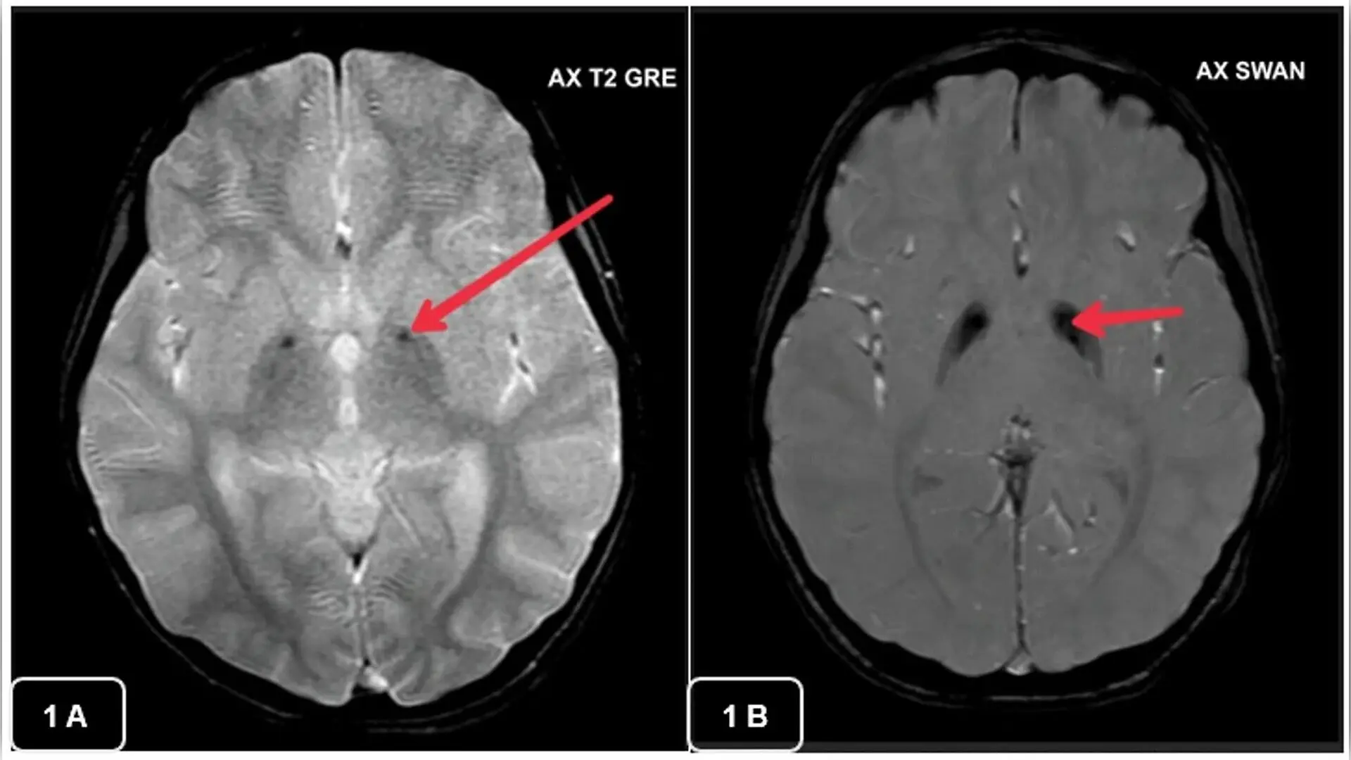
Four children in South India with rare neurodegenerative conditions experienced diagnostic delays before genetic testing revealed specific gene mutations tied to brain iron buildup. The young patients displayed severe motor difficulties and movement disorders that initially baffled clinicians or mimicked other conditions like Wilson disease.
Neurodegeneration with brain iron accumulation represents an ultra-rare group of inherited disorders where iron builds up destructively inside deep brain structures called the basal ganglia. These regions handle movement control, and excess iron triggers toxic oxidative stress and cell death. The researchers detailed their findings published in the Journal of Medical Case Reports, shedding light on the diagnostic hurdles families face in resource-limited settings.
What Specific Mutations Drive These Rare Disorders
The medical team evaluated two distinct subtypes among the four pediatric patients. Two unrelated young girls, aged 4 and 6 years old, presented with early-onset muscle stiffness, delayed development, and unsteadiness. Brain scans showed the classic eye-of-the-tiger sign, which features a bright spot surrounded by dark, iron-laden tissue in a brain region called the globus pallidus. Whole-exome sequencing, a laboratory method that reads all protein-coding genes, confirmed pathogenic variants in the PANK2 gene, establishing a diagnosis of pantothenate kinase-associated neurodegeneration.
The other two patients were 16-year-old cousins from a consanguineous family who developed walking difficulties and slurred speech during mid-childhood. Their brain magnetic resonance imaging scans displayed hyperintense streaking rather than the classic tiger eye pattern. Genetic analysis identified a variant in the C19orf12 gene, confirming mitochondrial membrane protein-associated neurodegeneration. Both teenagers suffered from progressive vision loss due to optic nerve atrophy alongside worsening muscle rigidity and abnormal posturing.
Why Brain Iron Accumulation Mimics Other Conditions
Recognizing these rare genetic conditions presents a significant clinical challenge because their early symptoms overlap heavily with more common neurological disorders. One of the 16-year-old patients was initially treated for neurological Wilson disease, a disorder of copper metabolism, because his brain scans and borderline laboratory tests muddied the clinical picture. He received zinc and D-penicillamine medications that ultimately failed to slow his neurological decline before genetic testing exposed the true molecular culprit.
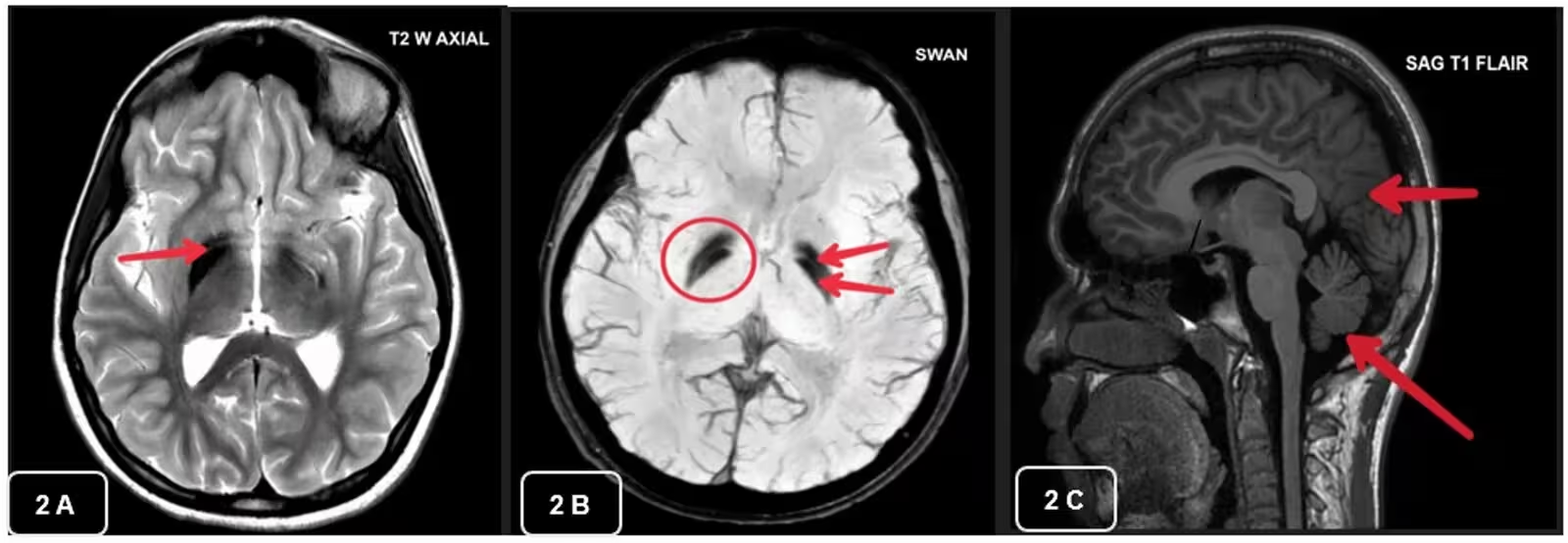
Financial constraints, the necessity of hospital admissions for sedated brain scans, and restricted access to advanced genomic panels historically delayed definitive answers for these families. Although next-generation sequencing costs have dropped, obtaining timely molecular tests remains a hurdle in regional pediatric centers.
What These Diagnostic Insights Mean for Future Care
No definitive cure currently exists for neurodegeneration with brain iron accumulation, so doctors rely on supportive therapies like physical therapy, speech therapy, and medications such as baclofen to manage painful muscle spasms. However, identifying the exact genetic subtype opens doors for family genetic counseling, accurate carrier testing, and potential future enrollment in clinical trials evaluating targeted therapies like brain-penetrant enzyme activators.
The authors concluded that integrating detailed neuroimaging patterns with whole-exome sequencing is essential for ending diagnostic odysseys. Expanding regional disease registries and broadening genomic screening access will help physicians catch these devastating conditions earlier and improve supportive care for affected children.
The research was published in Journal of medical case reports on November 13, 2026.
This article has been fact checked for accuracy, with information verified against reputable sources. Learn more about us and our editorial process.
Last reviewed on .
Article history
- Latest version
Reference(s)
- Mathew, Nelia., et al. “The clinical, radiological, and genetic features of Pantothenate Kinase-Associated Neurodegeneration and Mitochondrial membrane Protein-Associated Neurodegeneration in Neurodegeneration with Brain Iron Accumulation: a case series.” Journal of Medical Case Reports, vol. 20, no. 1, November 13, 2026 Springer Science and Business Media LLC, doi: 10.1186/s13256-026-06790-6. <https://doi.org/10.1186/s13256-026-06790-6>.
Cite this page:
- Posted by Faisal Qureshi