Parasites Hijack Host Cellular Architecture to Fuel Their Replication
Genetics

Parasites Hijack Host Cellular Architecture to Fuel Their Replication

A newly identified host protein response helps Leishmania remodel macrophage mitochondria to establish infection.
By Faisal Qureshi
Published:
Email this Article
Parasites Hijack Host Cellular Architecture to Fuel Their Replication
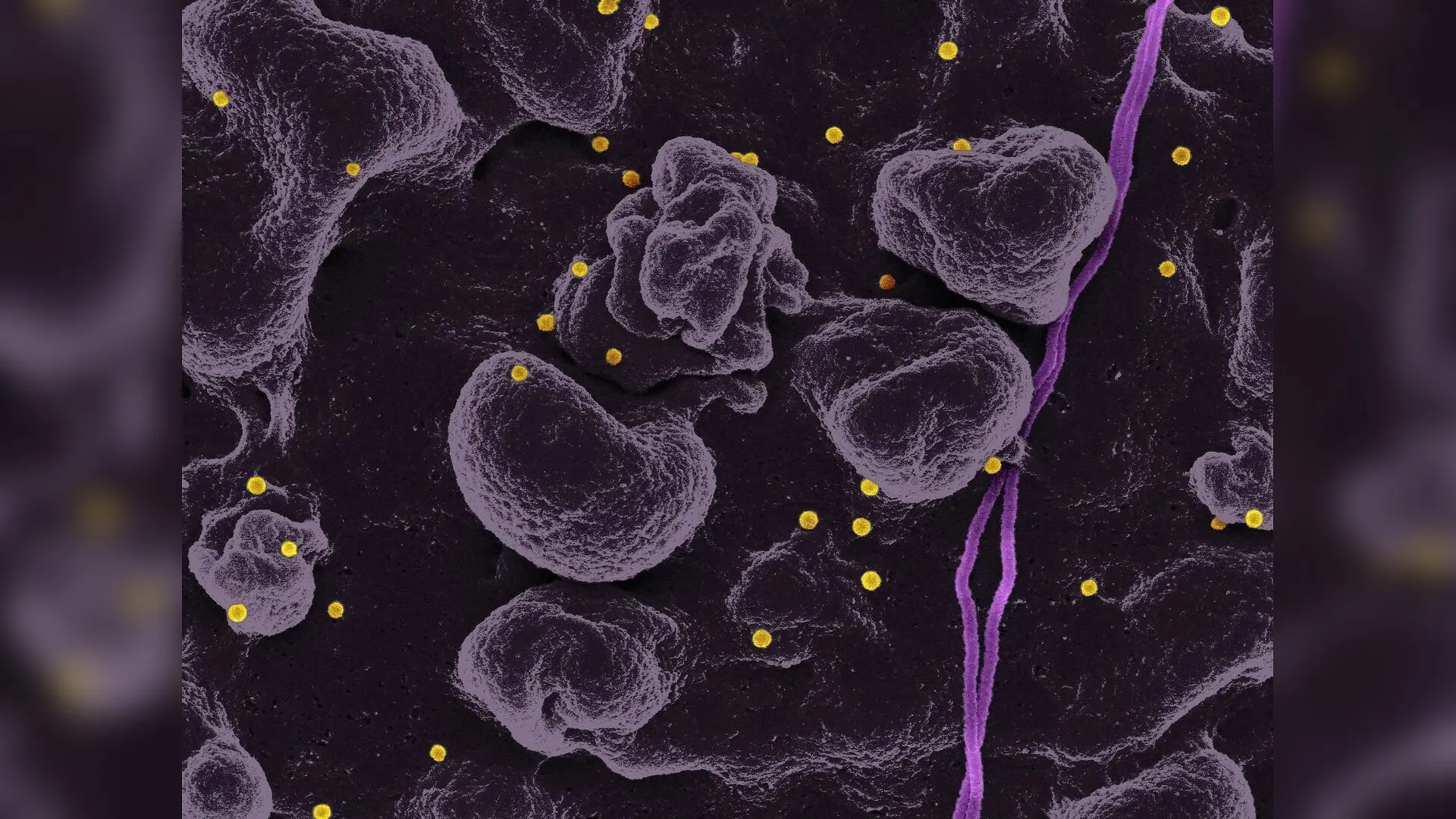
Photo by National Institute of Allergy and Infectious Diseases on Unsplash

Single-celled parasites responsible for leishmaniasis hijack a host cell structure called the endoplasmic reticulum to help them multiply inside immune cells. When researchers analyzed mouse macrophages, they discovered that a specific host protein helps the parasite remodel cellular power plants to fuel its own growth.

How Parasites Target Cellular Architecture

Leishmania parasites survive inside mammalian macrophages by taking up residence within specialized compartments known as parasitophorous vacuoles. These protective bubbles interact extensively with the macrophage endoplasmic reticulum, a sprawling network of membranes responsible for lipid synthesis and protein folding. Charles-Antoine Boyer and colleagues investigated whether the structural shape of this host network influences infection, publishing their findings in PLOS Pathogens.

The team focused on two major proteins that maintain endoplasmic reticulum architecture: CLIMP-63, which stabilizes flat membrane sheets, and RTN4, which shapes curved tubules. While RTN4 distribution remained unchanged during infection, the researchers observed a striking redistribution of CLIMP-63 away from its normal cellular locations and toward the parasite-containing vacuoles.
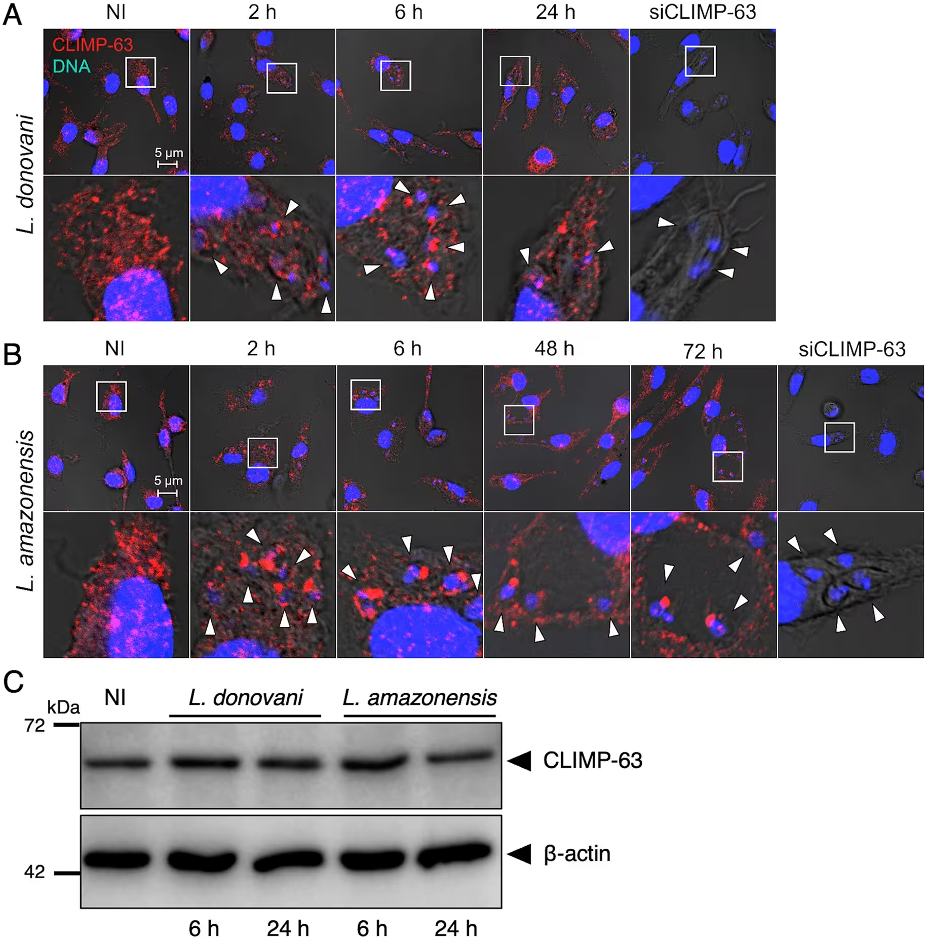
Fig 1: Leishmania induces the redistribution of CLIMP-63. BMM were infected or not (NI) with either (A) L. donovani or (B) L. amazonensis metacyclic promastigotes and at the indicated times points post-infection, the distribution of CLIMP-63 (red) was assessed by confocal immunofluorescence microscopy. DNA is in blue. White arrowheads denote internalized parasites. 5X-enlarged insets of representative localization of CLIMP-63 are shown. Controls for the specificity of the anti-CLIMP-63 antibody were performed using BMM pretreated with siRNA to CLIMP-63 and infected for 6 h with either L. donovani or L. amazonensis metacyclic promastigotes. Results are representative of at least three independent experiments. (C) Lysates of uninfected BMM (NI) and of BMM infected with either L. donovani or L. amazonensis metacyclic promastigotes were prepared at the indicated time points in lysis buffer containing 10 mM 1,10-phenanthroline. Levels and integrity of CLIMP-63 were assessed by Western blot analysi
Leishmania induces the redistribution of CLIMP-63. BMM were infected or not (NI) with either (A) L. donovani or (B) L. amazonensis metacyclic promastigotes and at the indicated times points post-infection, the distribution of CLIMP-63 (red) was assessed by confocal immunofluorescence microscopy. DNA is in blue. White arrowheads denote internalized parasites. 5X-enlarged insets of representative localization of CLIMP-63 are shown. Controls for the specificity of the anti-CLIMP-63 antibody were performed using BMM pretreated with siRNA to CLIMP-63 and infected for 6 h with either L. donovani or L. amazonensis metacyclic promastigotes. Results are representative of at least three independent experiments. (C) Lysates of uninfected BMM (NI) and of BMM infected with either L. donovani or L. amazonensis metacyclic promastigotes were prepared at the indicated time points in lysis buffer containing 10 mM 1,10-phenanthroline. Levels and integrity of CLIMP-63 were assessed by Western blot analysi Credit: Charles-Antoine Boyer et al. / PLOS Pathogens

The Role of Surface Glycolipids in Protein Hijacking

Further analysis revealed that this protein relocation depends entirely on a surface molecule called lipophosphoglycan, an abundant glycolipid covering the parasite. When macrophages were infected with mutant parasites lacking this specific glycolipid, CLIMP-63 failed to accumulate around the vacuoles. This chemical signal allows the pathogen to actively reorder host cellular components to suit its developmental needs.

Once redirected to the vacuole surface, CLIMP-63 showed an increased association with VDAC2, a protein located at contact sites between the endoplasmic reticulum and mitochondria. Mitochondria serve as the energy-producing power plants of the cell, and Leishmania is known to manipulate these organelles to secure nutrients.
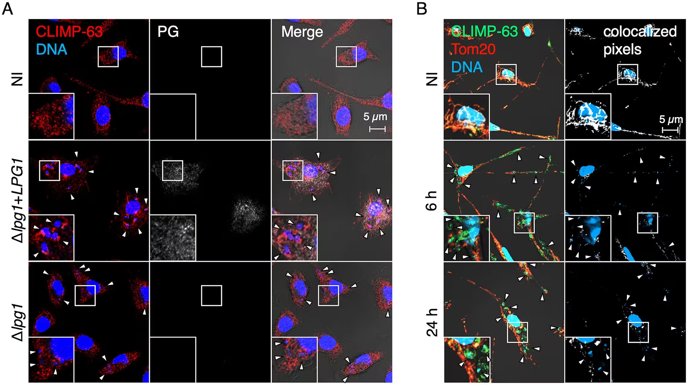
Fig 2: Redistribution of CLIMP-63 is LPG-dependent and coincides with its dissociation from the mitochondrial network. (A) BMM were infected or not (NI) with either Δlpg1 or Δlpg1 + LPG1 L. donovani metacyclic promastigotes and at the indicated times points the distribution of CLIMP-63 (red) was assessed by confocal immunofluorescence microscopy. DNA is in blue. White arrowheads denote internalized parasites. (B) BMM were infected with L. donovani metacyclic promastigotes and at the indicated times points the distribution and colocalization of CLIMP-63 (green) and Tom20 (red) were assessed by confocal immunofluorescence microscopy. Merge images and colocalized pixels (white) are shown. DNA is in blue. White arrowheads denote internalized parasites. Results are representative of at least three independent experiments.
Redistribution of CLIMP-63 is LPG-dependent and coincides with its dissociation from the mitochondrial network. (A) BMM were infected or not (NI) with either Δlpg1 or Δlpg1 + LPG1 L. donovani metacyclic promastigotes and at the indicated times points the distribution of CLIMP-63 (red) was assessed by confocal immunofluorescence microscopy. DNA is in blue. White arrowheads denote internalized parasites. (B) BMM were infected with L. donovani metacyclic promastigotes and at the indicated times points the distribution and colocalization of CLIMP-63 (green) and Tom20 (red) were assessed by confocal immunofluorescence microscopy. Merge images and colocalized pixels (white) are shown. DNA is in blue. White arrowheads denote internalized parasites. Results are representative of at least three independent experiments. Credit: Charles-Antoine Boyer et al. / PLOS Pathogens

Rescuing Parasite Replication Through Metabolic Boosts

To determine whether CLIMP-63 is necessary for infection, the investigators used small interfering RNA molecules to deplete the protein in mouse bone marrow-derived macrophages. Without CLIMP-63, parasite burdens dropped by about 30 percent during the first 24 hours and DNA replication among the amastigotes fell by up to half, showing that the host factor aids successful colonization.

Loss of CLIMP-63 also prevented the normal infection-induced increase in mitochondrial DNA synthesis, cristae remodeling, and oxygen consumption. When the team treated the depleted cells with AICAR, a chemical compound that stimulates mitochondrial biogenesis, the treatment partially restored the ability of the parasites to replicate. This experimental rescue demonstrates that the endoplasmic reticulum protein supports infection primarily by enabling essential metabolic changes in host mitochondria.

The research was published in PLOS Pathogens on November 18, 2026.

Fact Checked

This article has been fact checked for accuracy, with information verified against reputable sources. Learn more about us and our editorial process.

Last reviewed on .

Article history

  • Latest version

Reference(s)

  1. Boyer, Charles-Antoine., et al. “Leishmania exploits the macrophage endoplasmic reticulum-shaping protein CLIMP-63 to modulate mitochondrial biogenesis and bioenergetics.” PLOS Pathogens, vol. 22, no. 11, November 18, 2026, pp. e1014100 Public Library of Science (PLoS), doi: 10.1371/journal.ppat.1014100. <https://doi.org/10.1371/journal.ppat.1014100>.

Cite this page:

Qureshi, Faisal. “Parasites Hijack Host Cellular Architecture to Fuel Their Replication.” BioScience. BioScience ISSN 2521-5760, 19 November 2026. <https://www.bioscience.com.pk/en/subject/genetics/leishmania-exploits-the-macrophage-endoplasmic-reticulum-shaping-protein-climp-63-to-modulate-mitochondrial-biogenesis-and-bioenergetics>. Qureshi, F. (2026, November 19). “Parasites Hijack Host Cellular Architecture to Fuel Their Replication.” BioScience. ISSN 2521-5760. Retrieved November 19, 2026 from https://www.bioscience.com.pk/en/subject/genetics/leishmania-exploits-the-macrophage-endoplasmic-reticulum-shaping-protein-climp-63-to-modulate-mitochondrial-biogenesis-and-bioenergetics Qureshi, Faisal. “Parasites Hijack Host Cellular Architecture to Fuel Their Replication.” BioScience. ISSN 2521-5760. https://www.bioscience.com.pk/en/subject/genetics/leishmania-exploits-the-macrophage-endoplasmic-reticulum-shaping-protein-climp-63-to-modulate-mitochondrial-biogenesis-and-bioenergetics (accessed November 19, 2026).
End of the article