New AI Method Reconstructs Hidden Tissue Growth and Cell Fates Across Time
A newly developed computational framework uses flow matching and optimal transport to interpolate missing developmental stages from static spatial transcriptomic snapshots.
A new machine learning method can predict how tissues grow and change shape over time by tracking single cells that cannot normally be followed continuously. The computational tool, named ST-FateFlow, reconstructs both gene activity and physical tissue architecture across unobserved time points by bridging static snapshots of developing and regenerating biological systems.
Because standard laboratory methods destroy tissue during profiling, scientists have long struggled to connect individual cells from one time point to the next. Researchers Zhe Yu, Xiaobo Sun, and Hao Wu designed their new framework, published in arXiv, to solve this matching problem using optimal transport mathematics, which calculates the most likely paths cells take as they differentiate and migrate.
How the Model Predicts Missing Biological Stages
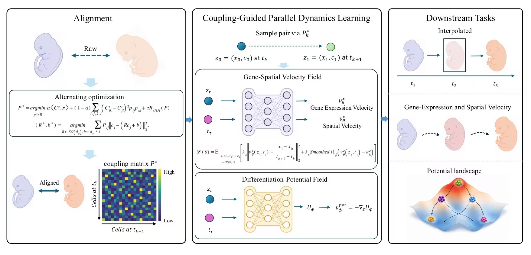
The framework operates by coupling adjacent spatial transcriptomic data snapshots, which measure both gene expression and physical coordinates in two or three dimensions. Instead of forcing a single mathematical rule on the data, the architecture separates the problem into two distinct tasks. One part of the network learns flexible velocity fields for gene expression and tissue deformation, allowing it to accurately interpolate missing intermediate states between observed time points. Another separate branch learns a time-conditioned potential function that organizes cell states along differentiation progressions from progenitor cells to fully specialized tissue types.
The authors evaluated the method across three large experimental datasets covering mouse embryonic development, planarian whole-body regeneration, and axolotl brain regeneration. In tests where researchers deliberately hid intermediate time points from the algorithm, the model successfully reconstructed both molecular distributions and tissue geometry with lower error rates than existing computational alternatives.
What the Potential Landscapes Reveal About Regeneration
When analyzing axolotl brain regeneration, the learned potential landscape correctly ordered known cell trajectories, showing that progenitor populations occupy higher potential states while specialized neurons sit at lower potential positions. By screening genes along these trajectories, the team identified potential-associated markers such as SFRP1, a modulator of Wnt signaling linked to neural progenitor maintenance, and CEBPD, a transcription factor involved in injury and inflammation responses.
The authors note that these statistical associations highlight candidate genes for cell-state progression rather than proving direct causal roles. They also emphasize that the learned potential represents a relative organization of cells rather than a physical energy or absolute fate probability. Future applications could help researchers model complex tissue remodeling in diverse developmental and disease contexts without requiring continuous live-cell tracking.
The study was posted to the arXiv preprint server on November 17, 2026.
This article has been fact checked for accuracy, with information verified against reputable sources. Learn more about us and our editorial process.
Last reviewed on .
Article history
- Latest version
Reference(s)
- Yu, Zhe. “ST-FateFlow: Continuous Gene-Spatial Reconstruction with Differentiation-Potential Learning from Time-Resolved Spatial Transcriptomics.” arXiv.org <http://arxiv.org/abs/2611.22256v1>.
Cite this page:
- Posted by Faisal Qureshi