Crucial Heart Receptor Deficiency Disrupts Atrial Development
Genetic models reveal that TIE1 and TIE2 work in synergy to build the heart’s upper chambers, uncovering potential roots of atrial cardiomyopathy.
When embryonic heart development goes awry, the delicate internal muscular networks of the chambers often fail to form properly, leading to congenital heart conditions. Researchers studying mouse models have discovered that a critical endothelial receptor protein known as TIE1 is essential for building the complex internal trabeculae inside the atria. The findings reveal that TIE1 and its partner TIE2 work together to regulate chamber morphogenesis, operating much more prominently in the upper heart chambers than in the lower ventricles.
Atrial cardiomyopathy involves structural and functional alterations in the heart’s upper chambers, yet the genetic blueprint driving atrial development has remained poorly understood compared to ventricular formation. To explore how chamber walls take shape, investigators examined published single-cell RNA sequencing datasets alongside bulk transcriptomic analyses of embryonic mouse hearts. The work is published in eLife, detailing how the genetic deletion of developmental regulators impacts cardiac architecture.
Differential Receptor Expression Shapes Atrial Architecture
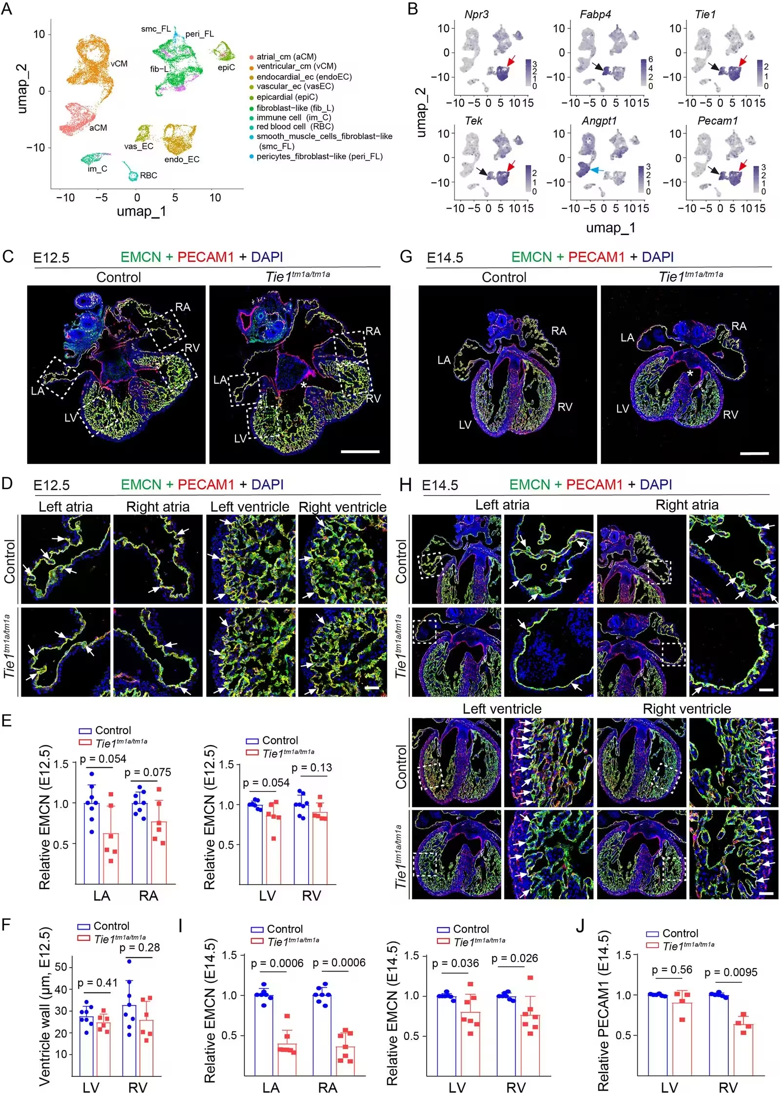
Single-cell transcriptomic profiling showed that both Tie1 and its homolog Tek, which encodes the TIE2 receptor, are expressed at significantly higher levels in atrial endocardial cells than in ventricular endocardial cells during embryonic development. Computational ligand-receptor analysis further indicated that ANGPT1-TIE2 signaling between atrial cardiomyocytes and endocardial cells is considerably stronger in the atria. When scientists analyzed mutant mouse lines lacking functional TIE1, they observed a striking absence of atrial trabeculae by embryonic day 14.5, whereas ventricular structures exhibited only sparse and milder defects.
Further investigations utilizing inducible endothelial knockout models demonstrated that losing TIE2 function similarly impairs atrial trabeculation within just 48 hours of deletion. Furthermore, combining a TIE1 deficiency with a heterozygous knockout of TIE2 resulted in severe developmental suppression in both atria and ventricles as early as embryonic day 10.5, highlighting a strong synergistic relationship between the two receptors. Transcriptomic profiling of these mutant hearts revealed a significant downregulation of key developmental genes, including Tek, Dll1, and Notch1, pointing toward disrupted endocardial-myocardial signaling pathways.
What These Findings Mean for Future Treatments
While mutations in the TIE1 gene have previously been identified in patients suffering from lymphedema, it remained unclear whether those individuals also experience underlying cardiac structural defects. The new genetic evidence bridges this gap by demonstrating that TIE1 and TIE2 are directly required for the endocardial-mediated organization of the atrial internal muscular network. Because defects in atrial trabeculation and remodeling can eventually lead to atrial dysfunction and heart failure, understanding these signaling axes provides a valuable foundation for future translational research into chamber-specific cardiomyopathies.
The authors note that fully mapping out the downstream signaling cascades will require isolating primary endocardial cells and performing high-resolution spatial transcriptomics on mutant embryos. Nevertheless, identifying the distinct vulnerabilities of atrial endocardium sheds light on why certain congenital heart defects manifest primarily in the upper chambers, offering fresh targets for diagnostic and therapeutic strategies.
The research was published in eLife on November 16, 2026.
This article has been fact checked for accuracy, with information verified against reputable sources. Learn more about us and our editorial process.
Last reviewed on .
Article history
- Latest version
Reference(s)
- Ding, Kai., et al. “Endocardial TIE1 synergizes with TIE2 to regulate the atrial internal muscular network assembly.” eLife, vol. 15, November 16, 2026 eLife Sciences Publications, Ltd, doi: 10.7554/eLife.111156. <https://doi.org/10.7554/eLife.111156>.
Cite this page:
- Posted by Elizabeth Taylor