Active Protein Machines Can Propel and Reshape Lipid Rafts
Chemistry

Active Protein Machines Can Propel and Reshape Lipid Rafts

Theoretical modeling shows that internal molecular motors create hydrodynamic stresses that push and twist cellular domains into unexpected trajectories.
By Anjali Singh
Published:
Email this Article
Active Protein Machines Can Propel and Reshape Lipid Rafts
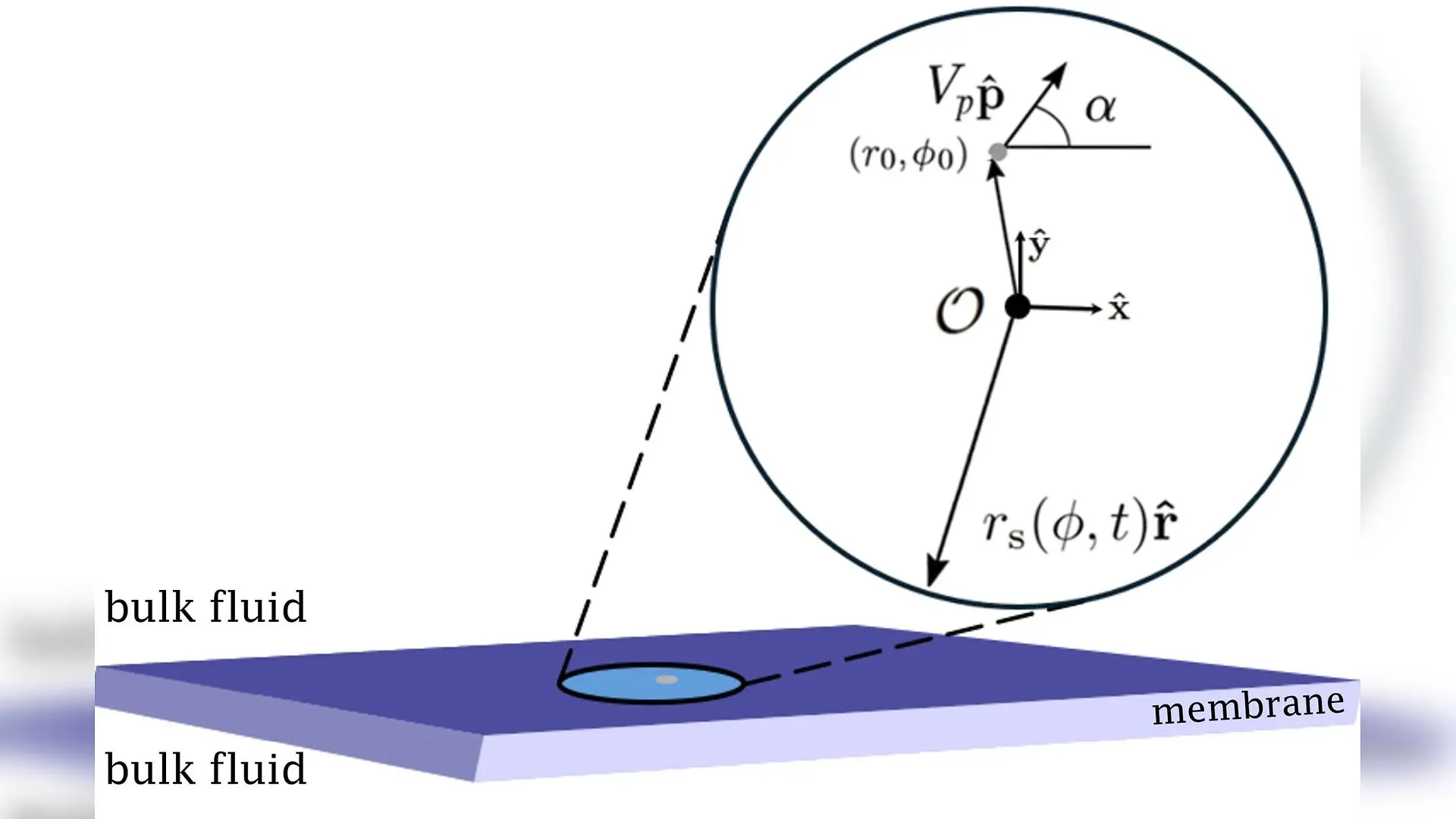
Problem setup: A quasi- circular domain of viscosity λ​ηm\lambda\eta_{m} in a planar membrane of viscosity ηm\eta_{m}. Inside the domain, an active particle located at x0=(r0,ϕ0)\textbf{x}_{0}=(r_{0},\phi_{0}) moves with active velocity VpV_{p} in direction 𝐩^{\bf\hat{p}}. The vector 𝐩^{\bf\hat{p}} forms an angle α\alpha with the x-axis. The domain boundary is free to evolve in response to the flow generated by the active particle and the domain shape is parametrized by rs​(ϕ,t)r_{\mathrm{s}}(\phi,t). Gonzalo Ferrandez Quinto et al. / arXiv

Biological cell membranes are fluid, nanometer-thin lipid sheets packed with specialized protein machinery, including tiny molecular motors and chemical pumps that constantly consume energy to perform mechanical work. Northwestern University researchers Gonzalo Ferrandez Quinto, Sho Kawakami, and Petia M. Vlahovska have investigated how these active molecular machines influence their immediate environment, revealing that force-free internal activities can actively propel and reshape specialized cholesterol-rich membrane domains known as lipid rafts.

The study, available on the arXiv preprint server, models active protein machinery as point singularities operating within a two-dimensional Stokes flow. Biological membranes are crowded environments where viscous forces dominate and inertia plays virtually no role. Under such physical conditions, any force exerted by a protein machine on the surrounding fluid is met by an equal and opposite reaction, meaning that internal biomolecules often behave dynamically as force dipoles or source dipoles.

How Internal Protein Pumps Drive Membrane Droplet Motion

The authors developed an analytical framework to track how active forces generated inside a circular membrane domain, such as a fluid lipid raft, translate into physical movement and boundary deformation. While traditional views often assume that lipid rafts drift passively through a membrane via simple thermal diffusion, this new work suggests that the internal churning of active enzymes creates localized hydrodynamic stresses that alter the shape and trajectory of the entire domain.
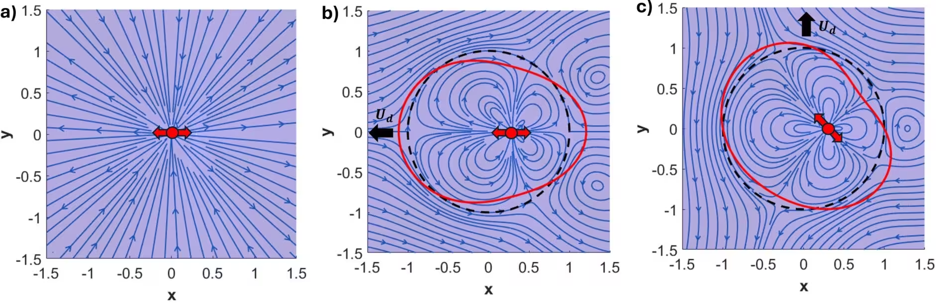
Figure 2: Flow of a stresslet in free space and confined inside a circular domain. (a) Flow about an unconfined stresslet placed at the origin. (b), (c) Flow created by a stresslet confined in a domain and located off-center at (r0,ϕ0)=(0.3,0)(r_{0},\phi_{0})=(0.3,0). The stresslet is tilted relative to the line connecting its position to the domain’s center, with angles α=0\alpha=0 and α=3​π/4\alpha=3\pi/4, respectively. In all plots C​a=0.04Ca=0.04 and λ=1\lambda=1. The red dot represents the location of the stresslet. The black line represents the undisturbed domain shape and the red line represents the equilibrium shape of the domain. The black arrow represents the direction of motion of the domain. For (b) it points to the left and for (c) it points upwards.
Flow of a stresslet in free space and confined inside a circular domain. (a) Flow about an unconfined stresslet placed at the origin. (b), (c) Flow created by a stresslet confined in a domain and located off-center at (r0,ϕ0)=(0.3,0)(r_{0},\phi_{0})=(0.3,0). The stresslet is tilted relative to the line connecting its position to the domain’s center, with angles α=0\alpha=0 and α=3​π/4\alpha=3\pi/4, respectively. In all plots C​a=0.04Ca=0.04 and λ=1\lambda=1. The red dot represents the location of the stresslet. The black line represents the undisturbed domain shape and the red line represents the equilibrium shape of the domain. The black arrow represents the direction of motion of the domain. For (b) it points to the left and for (c) it points upwards. Credit: Gonzalo Ferrandez Quinto et al. / arXiv

When examining a source dipole, which creates an irrotational fluid flow, the researchers found that the domain translates directly along the dipole axis at a velocity independent of the protein’s exact position within the domain. By contrast, a stresslet, which represents a force-free shear stress producer, generates a far more complex response. A stresslet does not propel the domain along its own axis of symmetry, meaning the resulting domain velocity depends sensitively on both where the protein is located and how it is oriented.

Confining Geometries Trigger Orbiting and Superdiffusion

The physical confinement of a circular droplet introduces important hydrodynamic constraints that change how active particles behave compared to free space. While an unconfined source dipole swims persistently in a straight line, confining it inside a fluid domain introduces vorticity and rotational flows that reorient the particle. Depending on the propulsion speed and the viscosity ratio between the domain and the surrounding membrane, the active particle can become trapped in stable fixed points or enter persistent orbital motion.

Furthermore, when a non-self-propelled stresslet undergoes random rotational diffusion inside the droplet, stochastic reorientation competes with self-induced hydrodynamic drift. The authors observed that the stresslet tends to align perpendicularly to its radial position vector, creating a directional bias that drives the host domain into a transient superdiffusive regime before thermal confinement and boundary repulsions take over at longer timescales.
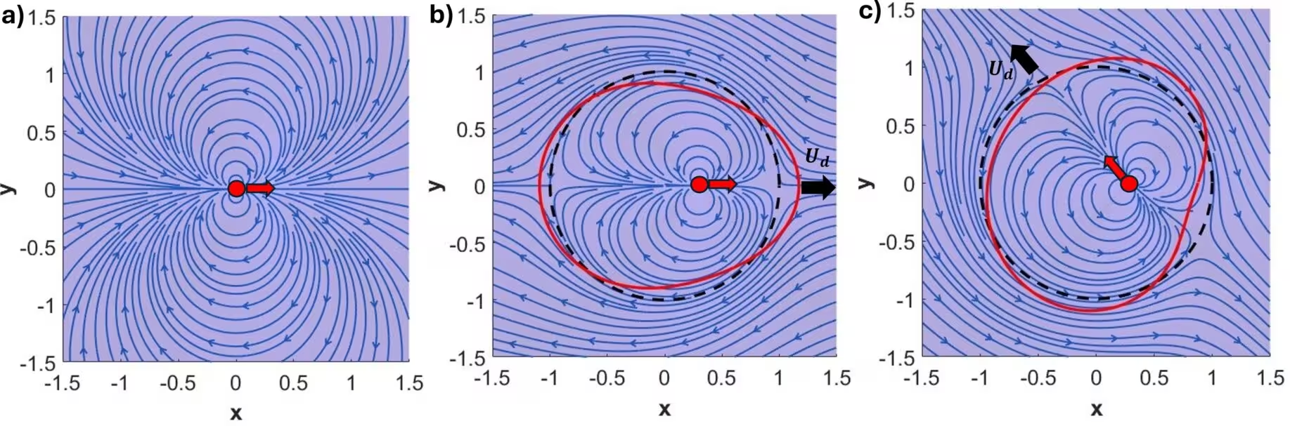
Figure 3: Flow due to a source dipole in free space and confined inside a circular domain. (a) Flow about an unconfined source dipole at the origin; all streamlines are closed. (b), (c) Flow created by a source dipole confined in a circular domain and located off-center at (x,y)=(0.3,0)(x,y)=(0.3,0). The dipole is tilted relative to the line connecting the dipole and the center of mass of the domain at angle α=0\alpha=0 and α=3​π/4\alpha=3\pi/4 correspondingly. The streamlines outside the domain are open, indicating domain motion. In all plots C​a=0.05Ca=0.05 and λ=1\lambda=1. The red dot represents the location of the dipole. The black line represents the undisturbed domain shape and the red line represents the equilibrium shape of the domain. The black arrow represents the migration direction of the domain; the fluid is being pumped in the opposite direction.
Flow due to a source dipole in free space and confined inside a circular domain. (a) Flow about an unconfined source dipole at the origin; all streamlines are closed. (b), (c) Flow created by a source dipole confined in a circular domain and located off-center at (x,y)=(0.3,0)(x,y)=(0.3,0). The dipole is tilted relative to the line connecting the dipole and the center of mass of the domain at angle α=0\alpha=0 and α=3​π/4\alpha=3\pi/4 correspondingly. The streamlines outside the domain are open, indicating domain motion. In all plots C​a=0.05Ca=0.05 and λ=1\lambda=1. The red dot represents the location of the dipole. The black line represents the undisturbed domain shape and the red line represents the equilibrium shape of the domain. The black arrow represents the migration direction of the domain; the fluid is being pumped in the opposite direction. Credit: Gonzalo Ferrandez Quinto et al. / arXiv

Physical Limitations and Future Biomimetic Applications

To make these complex equations solvable, the mathematical treatment relies on a regular perturbation analysis valid for small capillary numbers, where active hydrodynamic stresses remain relatively weak compared to the line tension holding the membrane domain circular. The model also assumes a flat, two-dimensional incompressible interface and treats the active protein machinery as point-like singularities.

The authors note that these theoretical findings provide a fundamental baseline for interpreting experiments on biomimetic membranes observed under optical microscopy. As artificial nanoengineering advances toward creating sophisticated self-propelled nanomotors, understanding how active particles interact with soft boundaries and fluid interfaces will be essential for mastering active transport at cellular scales.

The study was posted to the arXiv preprint server on November 19, 2026.

Fact Checked

This article has been fact checked for accuracy, with information verified against reputable sources. Learn more about us and our editorial process.

Last reviewed on .

Article history

  • Latest version

Reference(s)

  1. Quinto, Gonzalo. “Activity-driven motion and deformation of a circular membrane domain.” arXiv.org <http://arxiv.org/abs/2611.26578v1>.

Cite this page:

Singh, Anjali. “Active Protein Machines Can Propel and Reshape Lipid Rafts.” BioScience. BioScience ISSN 2521-5760, 20 November 2026. <https://www.bioscience.com.pk/en/subject/chemistry/activity-driven-motion-and-deformation-of-a-circular-membrane-domain>. Singh, A. (2026, November 20). “Active Protein Machines Can Propel and Reshape Lipid Rafts.” BioScience. ISSN 2521-5760. Retrieved November 20, 2026 from https://www.bioscience.com.pk/en/subject/chemistry/activity-driven-motion-and-deformation-of-a-circular-membrane-domain Singh, Anjali. “Active Protein Machines Can Propel and Reshape Lipid Rafts.” BioScience. ISSN 2521-5760. https://www.bioscience.com.pk/en/subject/chemistry/activity-driven-motion-and-deformation-of-a-circular-membrane-domain (accessed November 20, 2026).
End of the article