Yeast Biosurfactants Trigger Severe Stress and Cell Death in Breast Cancer Models
Biotechnology

Yeast Biosurfactants Trigger Severe Stress and Cell Death in Breast Cancer Models

Laboratory findings show that microbial glycolipids induce S-phase arrest and membrane disruption in human breast adenocarcinoma cells.
By Robert Brown
Published:
Email this Article
Yeast Biosurfactants Trigger Severe Stress and Cell Death in Breast Cancer Models
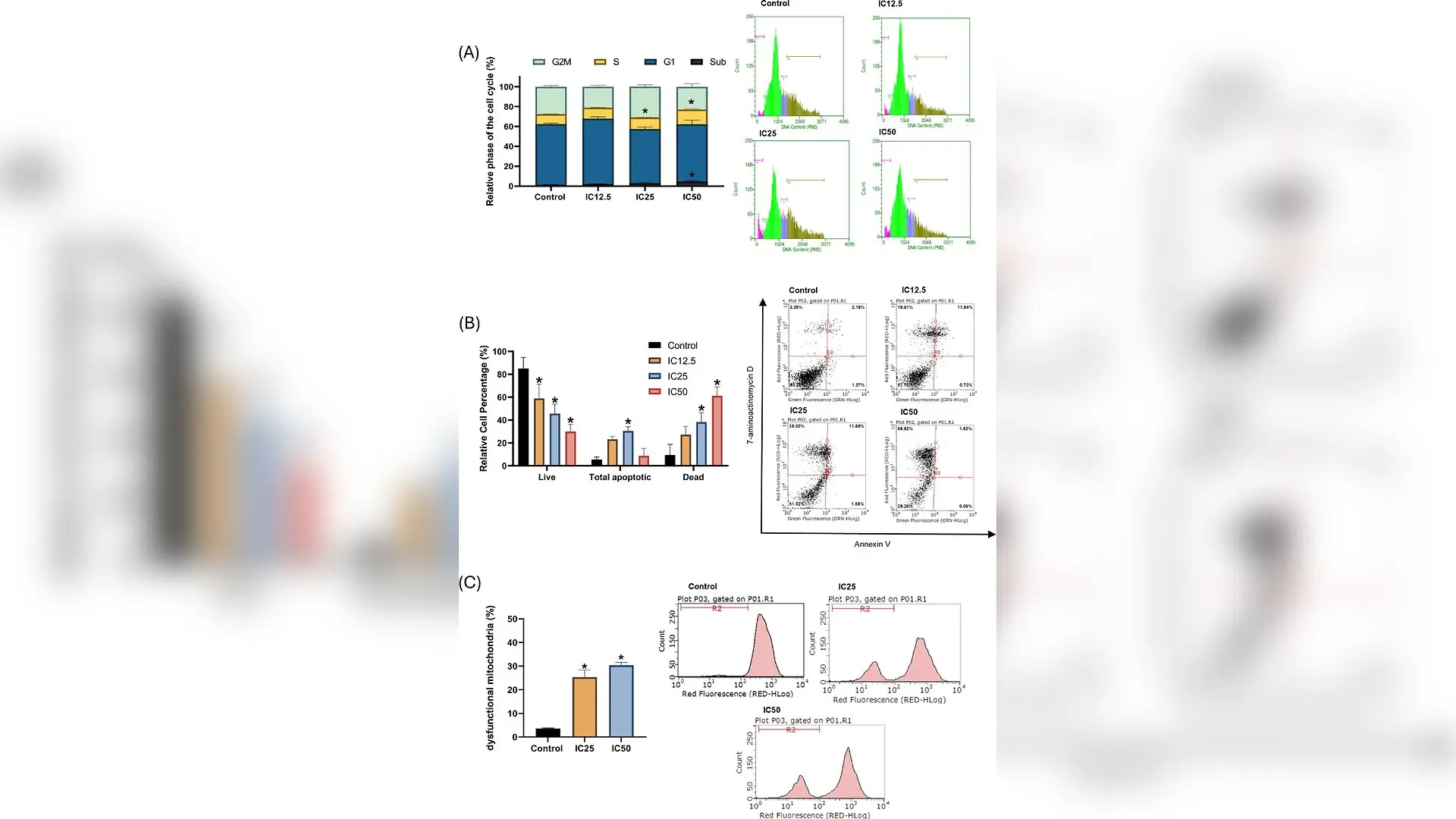
Flow cytometry analysis of cell cycle progression, cell death, and mitochondrial membrane potential in MCF-7 cells. Cells were evaluated after 24 h of exposure to sophorolipids at IC12.5, IC25, and IC50 concentrations. A Percentage distribution of cell cycle phases determined by DNA content quantification using propidium iodide (PI). The treatment induced a concentration-dependent accumulation of cells in the S phase (cell cycle arrest). B Proportion of viable, apoptotic, and necrosis-like dead cells obtained by Annexin V and 7-AAD dual labeling. A significant reduction in cell viability and a predominance of necrosis-like cell death are observed at higher concentrations. C Assessment of mitochondrial membrane potential (\documentclass[12pt]{minimal} \usepackage{amsmath} \usepackage{wasysym} \usepackage{amsfonts} \usepackage{amssymb} \usepackage{amsbsy} \usepackage{mathrsfs} \usepackage{upgreek} \setlength{\oddsidemargin}{-69pt} \begin{document}$$\:\varDelta\:\varPsi\:m)\:$$\end{docume Luiz Henrique Santana Martins et al. / 3 Biotech

Yeast-derived biosurfactants can dismantle human breast cancer cells in laboratory tests by triggering severe cellular stress and membrane disruption, according to a recent study. Researchers investigated how a mixture of natural glycolipids impacts MCF-7 breast adenocarcinoma cells, revealing a potent cytotoxic effect that halts cell growth and damages vital internal structures.

How Biosurfactants Target Breast Cancer Cells

Breast cancer remains the most frequently diagnosed malignant neoplasm among women worldwide, accounting for roughly 2.3 million new cases and 670,000 deaths annually. While modern treatments continue to advance, persistent medical challenges and regional health disparities drive the search for safer, more effective therapeutic alternatives derived from natural sources.

In this laboratory investigation published in 3 Biotech, a research team led by scientists from the State University of Londrina in Brazil examined the precise mechanisms by which microbial glycolipids affect cancerous cells. The team tested a compound mixture containing 63.15% lactonic forms and 36.85% acidic forms, which are secondary metabolites produced by the non-pathogenic yeast Starmerella bombicola.

What Happens When Cancer Cells Meet Yeast Glycolipids

When exposed to these microbial molecules for 24 hours, human breast cancer cells experienced a dose-dependent reduction in cell viability. The half-maximal inhibitory concentration, representing the dose required to reduce cell viability by 50 percent, was measured at 82.54 micrograms per milliliter. Phase-contrast microscopy and fluorescence staining revealed severe morphological alterations, including cell swelling, loss of adhesion, and compromised plasma membrane integrity.
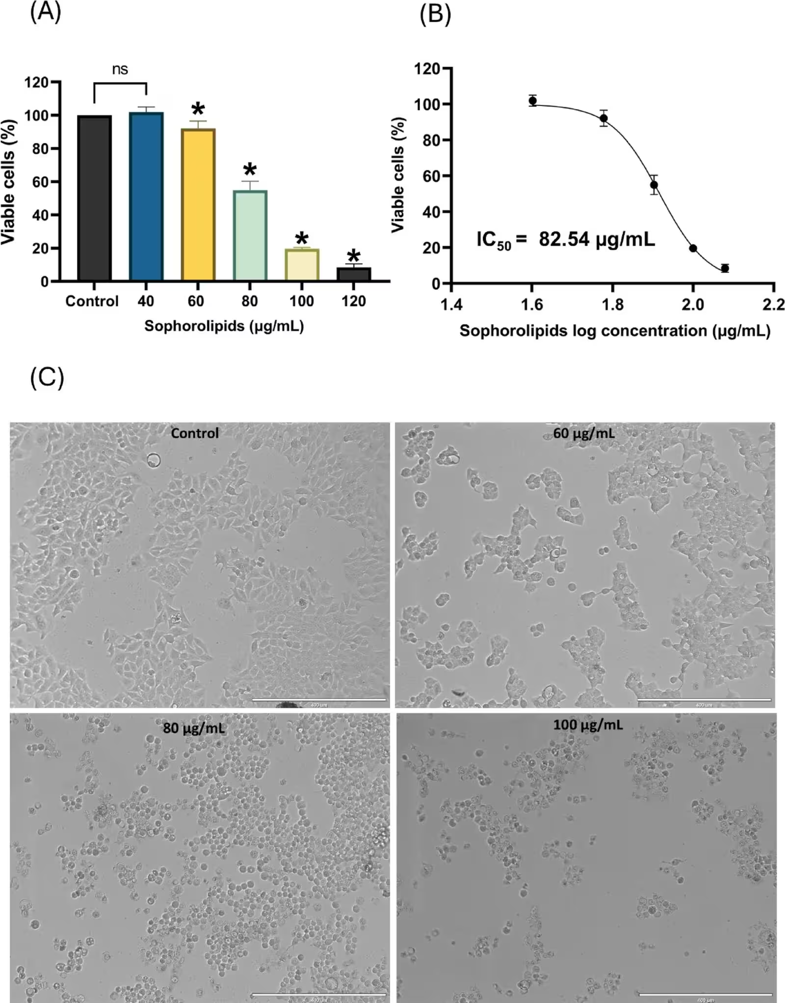
Fig. 1: Cytotoxic effect of sophorolipids on the mammary adenocarcinoma cell line (MCF-7). A Cell viability determined by the Resazurin assay after 24 h of exposure to various concentrations of sophorolipids (40–120 µg/mL), Doxorubicin was used as a positive control (see Supplementary Data). B Non-linear regression curve (dose-response) used to calculate the half-maximal inhibitory concentration IC50 and the IC25 and IC12.5 values. C Representative phase-contrast microscopy photomicrographs of MCF-7 cells after 24 h of treatment. The images highlight morphological alterations, including cell swelling, loss of morphology, reduced confluency, and cell detachment associated with cell death at higher concentrations. Scale bar 400 μm. Data are expressed as the mean ± standard deviation of three independent experimental replicates. Asterisks (*) indicate statistically significant differences compared to the control (p < 0.05) as determined by one-way Analysis of Variance (ANOVA) followed by Dunnett’
Cytotoxic effect of sophorolipids on the mammary adenocarcinoma cell line (MCF-7). A Cell viability determined by the Resazurin assay after 24 h of exposure to various concentrations of sophorolipids (40–120 µg/mL), Doxorubicin was used as a positive control (see Supplementary Data). B Non-linear regression curve (dose-response) used to calculate the half-maximal inhibitory concentration IC50 and the IC25 and IC12.5 values. C Representative phase-contrast microscopy photomicrographs of MCF-7 cells after 24 h of treatment. The images highlight morphological alterations, including cell swelling, loss of morphology, reduced confluency, and cell detachment associated with cell death at higher concentrations. Scale bar 400 μm. Data are expressed as the mean ± standard deviation of three independent experimental replicates. Asterisks (*) indicate statistically significant differences compared to the control (p < 0.05) as determined by one-way Analysis of Variance (ANOVA) followed by Dunnett’ Credit: Luiz Henrique Santana Martins et al. / 3 Biotech

Flow cytometry analysis further showed that the treatment caused cells to arrest in the S phase of the cell cycle, preventing normal division. Additionally, researchers observed significant mitochondrial membrane depolarization, indicating that the powerhouses of the cells were failing under metabolic strain.

Unraveling the Molecular Stress Pathways

At the transcriptional level, reverse transcription quantitative PCR demonstrated the overexpression of several key stress-response genes, including CDKN1A, GADD45A, ERN1, and BBC3. The upregulation of ERN1 points to endoplasmic reticulum stress, a state where cellular protein folding machinery becomes overwhelmed. Meanwhile, the increase in BBC3 activity promotes mitochondrial membrane permeability, compounding the cellular damage.
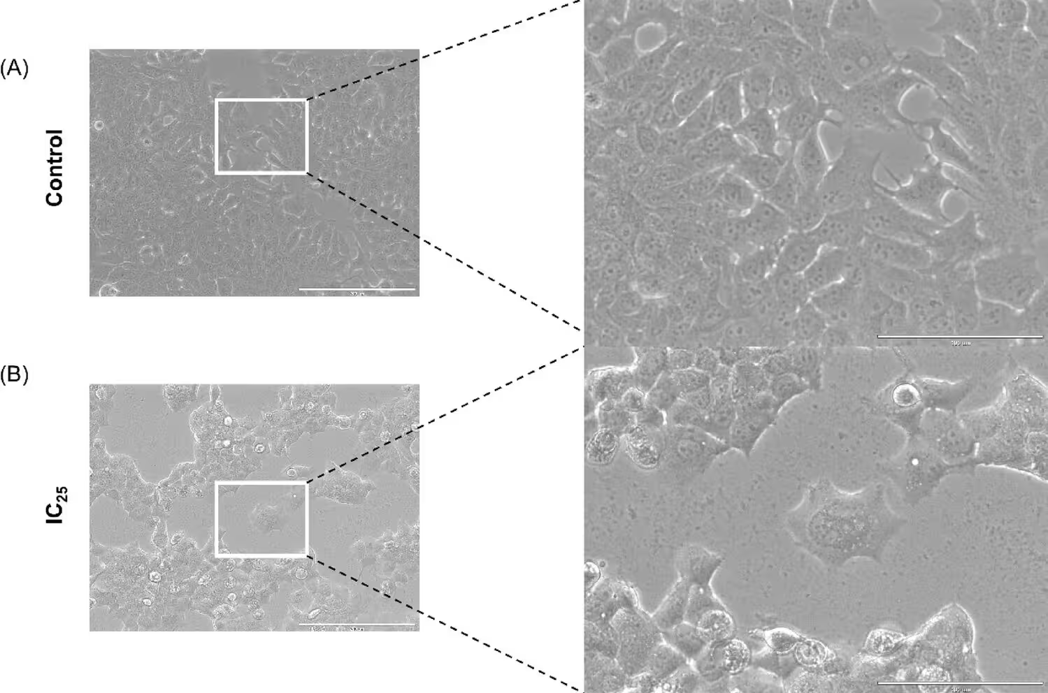
Fig. 2: Morphological analysis of MCF-7 cells via phase-contrast microscopy. A Untreated control group after 24 h of incubation, displaying typical epithelial-like morphology, high cell confluency, and intact cell-to-cell adhesion. B MCF-7 cells treated with the IC25 of sophorolipids (70.90 µg/mL) for 24 h. Notable morphological alterations include significant cell swelling, loss of characteristic polygonal shape, and a visible reduction in cell density compared to the control
Morphological analysis of MCF-7 cells via phase-contrast microscopy. A Untreated control group after 24 h of incubation, displaying typical epithelial-like morphology, high cell confluency, and intact cell-to-cell adhesion. B MCF-7 cells treated with the IC25 of sophorolipids (70.90 µg/mL) for 24 h. Notable morphological alterations include significant cell swelling, loss of characteristic polygonal shape, and a visible reduction in cell density compared to the control Credit: Luiz Henrique Santana Martins et al. / 3 Biotech

Interestingly, while the intrinsic apoptotic pathway was engaged, the cells predominantly underwent necrosis-like cell death, reaching 59.31 percent at the highest concentration tested. The authors note that the MCF-7 cell line naturally lacks functional caspase-3 due to a genetic deletion, which prevents the completion of classical programmed cell death. Instead, the amphiphilic nature of the glycolipids causes direct physical damage to the cell membrane, accompanied by the downregulation of structural and metabolic genes such as ACTB and GAPDH.

Next Steps for Natural Antiproliferative Therapies

These findings position microbial biosurfactants as promising candidates for the development of alternative antiproliferative therapies. However, the study authors emphasize several important limitations that must be addressed in future research. The experiments were conducted exclusively on a single in vitro cell line with specific genetic traits, and the use of an unseparated mixture of lactonic and acidic isoforms makes it impossible to isolate the exact contribution of each structural form.

Subsequent investigations will need to evaluate isolated fractions individually, incorporate non-malignant control lines, and extend the testing into complex in vivo models. These steps are necessary to fully understand the safety, efficacy, and clinical potential of these yeast-derived compounds before they can be considered for medical applications.

The research was published in 3 Biotech on November 1, 2026.

Fact Checked

This article has been fact checked for accuracy, with information verified against reputable sources. Learn more about us and our editorial process.

Last reviewed on .

Article history

  • Latest version

Reference(s)

  1. Martins, Luiz Henrique Santana., et al. “Investigation of the molecular mechanisms of action of sophorolipids in MCF-7 cells: therapeutic potential against breast cancer.” 3 Biotech, vol. 16, no. 11, October 23, 2026 Springer Science and Business Media LLC, doi: 10.1007/s13205-026-05096-0. <https://doi.org/10.1007/s13205-026-05096-0>.

Cite this page:

Brown, Robert. “Yeast Biosurfactants Trigger Severe Stress and Cell Death in Breast Cancer Models.” BioScience. BioScience ISSN 2521-5760, 27 October 2026. <https://www.bioscience.com.pk/en/subject/biotechnology/investigation-of-the-molecular-mechanisms-of-action-of-sophorolipids-in-mcf-7-cells-therapeutic-potential-against-breast-cancer>. Brown, R. (2026, October 27). “Yeast Biosurfactants Trigger Severe Stress and Cell Death in Breast Cancer Models.” BioScience. ISSN 2521-5760. Retrieved October 27, 2026 from https://www.bioscience.com.pk/en/subject/biotechnology/investigation-of-the-molecular-mechanisms-of-action-of-sophorolipids-in-mcf-7-cells-therapeutic-potential-against-breast-cancer Brown, Robert. “Yeast Biosurfactants Trigger Severe Stress and Cell Death in Breast Cancer Models.” BioScience. ISSN 2521-5760. https://www.bioscience.com.pk/en/subject/biotechnology/investigation-of-the-molecular-mechanisms-of-action-of-sophorolipids-in-mcf-7-cells-therapeutic-potential-against-breast-cancer (accessed October 27, 2026).
End of the article