Targeted Drug Halts Dangerous Aortic Aneurysm Growth in Preclinical Study
Biotechnology

Targeted Drug Halts Dangerous Aortic Aneurysm Growth in Preclinical Study

Researchers find that selective integrin inhibition blocks destructive cellular pathways in both genetic and sporadic forms of the cardiovascular disease.
By Rohan Kumar
Published:
Email this Article
Targeted Drug Halts Dangerous Aortic Aneurysm Growth in Preclinical Study
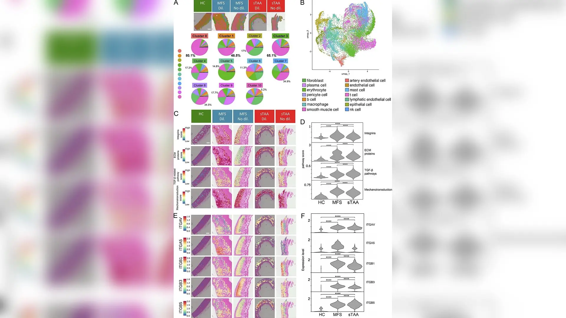
Transcription levels of genes encoding ECM components, TGF-β signalling mediators, mechanotransduction proteins, and integrins are higher in TAA of MFS and non-syndromic (sTAA) patients than in HC aortas. (A, C, and E) Spatial transcriptomics analysis on thoracic aortas from HC subjects and patients with MFS or sTAA, both in dilated (Dil.) and non-dilated (No dil.) aneurysmal regions. Scale bar = 1 mm. Aortic layers are displayed across all samples, from left to right, as intima, media, and adventitia. Images in A represent spatial spots. These spots have been detailed in pie charts, which summarize the aortic cell types defined by similar transcriptomic clusters. scRNA-seq data from the Tabula Sapiens vascular dataset (https://tabula-sapiens-portal.ds.czbiohub.org) have been used to perform the deconvolution and to attribute cellular composition to the spatial spots. The clusters highlighted (clusters 0, 1, and 3, in bold) contain the highest number of cells with transcriptional featu Sara Rega et al. / The Journal of experimental medicine

A targeted drug originally investigated for cancer therapy could halt the life-threatening ballooning of the body’s main artery by shutting down specific cellular receptors. Thoracic aortic aneurysm remains a major global health threat that typically requires major surgery once the vessel dilates past a dangerous threshold, because current medicines only offer palliative relief.

To investigate why these vessel walls weaken, a multi-institutional team of European researchers mapped spatial gene activity inside human aortic tissues. Their findings, published in the Journal of Experimental Medicine, reveal that transmembrane proteins called integrins containing the alpha-V subunit act as critical molecular switches driving destructive scarring and tissue degradation.

Mapping the Cellular Drivers of Vessel Weakening

The research team analyzed human tissue samples taken from healthy organ donors alongside specimens from patients undergoing surgical repair for sporadic thoracic aortic aneurysms and syndromic cases linked to Marfan syndrome. Using high-resolution spatial transcriptomics, the investigators mapped thousands of individual gene expressions across different layers of the aortic wall.
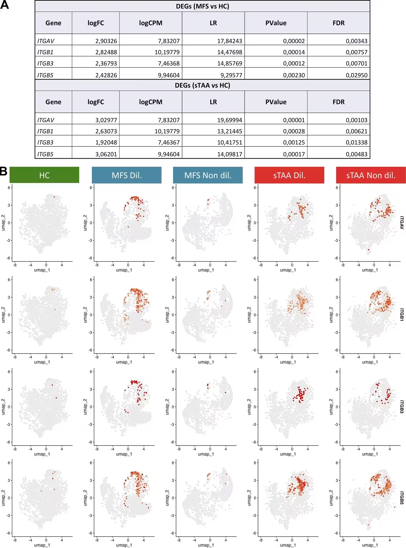
Figure S1: Integrin-related genes are predominantly expressed in VSMCs from MFS and sTAA aortas . (A) Tables reporting DEGs related to integrins obtained from spatial transcriptomics analysis data. (B) UMAP-based feature plots of the integrin genes (ITGAV, first row; ITGB1, second row; ITGB3, third row; ITGB5, fourth row). The gene expression normalized levels in VSMC are displayed with a shades-of-red color scale, from dark red (single-cell transform, SCT > 3.0, more highly expressed) to light red (SCT < 3.0, less expressed).
Integrin-related genes are predominantly expressed in VSMCs from MFS and sTAA aortas . (A) Tables reporting DEGs related to integrins obtained from spatial transcriptomics analysis data. (B) UMAP-based feature plots of the integrin genes (ITGAV, first row; ITGB1, second row; ITGB3, third row; ITGB5, fourth row). The gene expression normalized levels in VSMC are displayed with a shades-of-red color scale, from dark red (single-cell transform, SCT > 3.0, more highly expressed) to light red (SCT < 3.0, less expressed). Credit: Sara Rega et al. / The Journal of experimental medicine

The analysis showed that genes encoding alpha-V integrins, collagen remodelling proteins, and signaling molecules associated with transforming growth factor-beta were heavily upregulated in patient tissues compared to healthy controls. These alterations were particularly concentrated at the interface between the middle layer of the aorta and its outer adventitia, specifically within dilated regions where smooth muscle cells had shifted from a normal contractile state to a destructive secretory profile.

Testing Selective Versus Broad Inhibitors in the Lab

Building on these molecular maps, the authors isolated vascular smooth muscle cells from patients and tested two different integrin-blocking drugs to see if they could reverse the pathological changes. One compound, GLPG0187, acts as a broad-spectrum inhibitor blocking multiple integrin types, while the other, Cilengitide, selectively targets specific alpha-V dimers including alpha-V beta-3 and alpha-V beta-5.

Laboratory assays demonstrated that both compounds successfully reduced the release of active transforming growth factor-beta in patient cells without causing cellular toxicity. However, Cilengitide proved notably superior in suppressing both canonical and non-canonical signaling cascades linked to fibrosis. Furthermore, when the team knocked down the gene encoding the alpha-V subunit using lentiviral particles, the pro-fibrotic signaling was similarly blunted, confirming that these specific receptors control the destructive pathways.
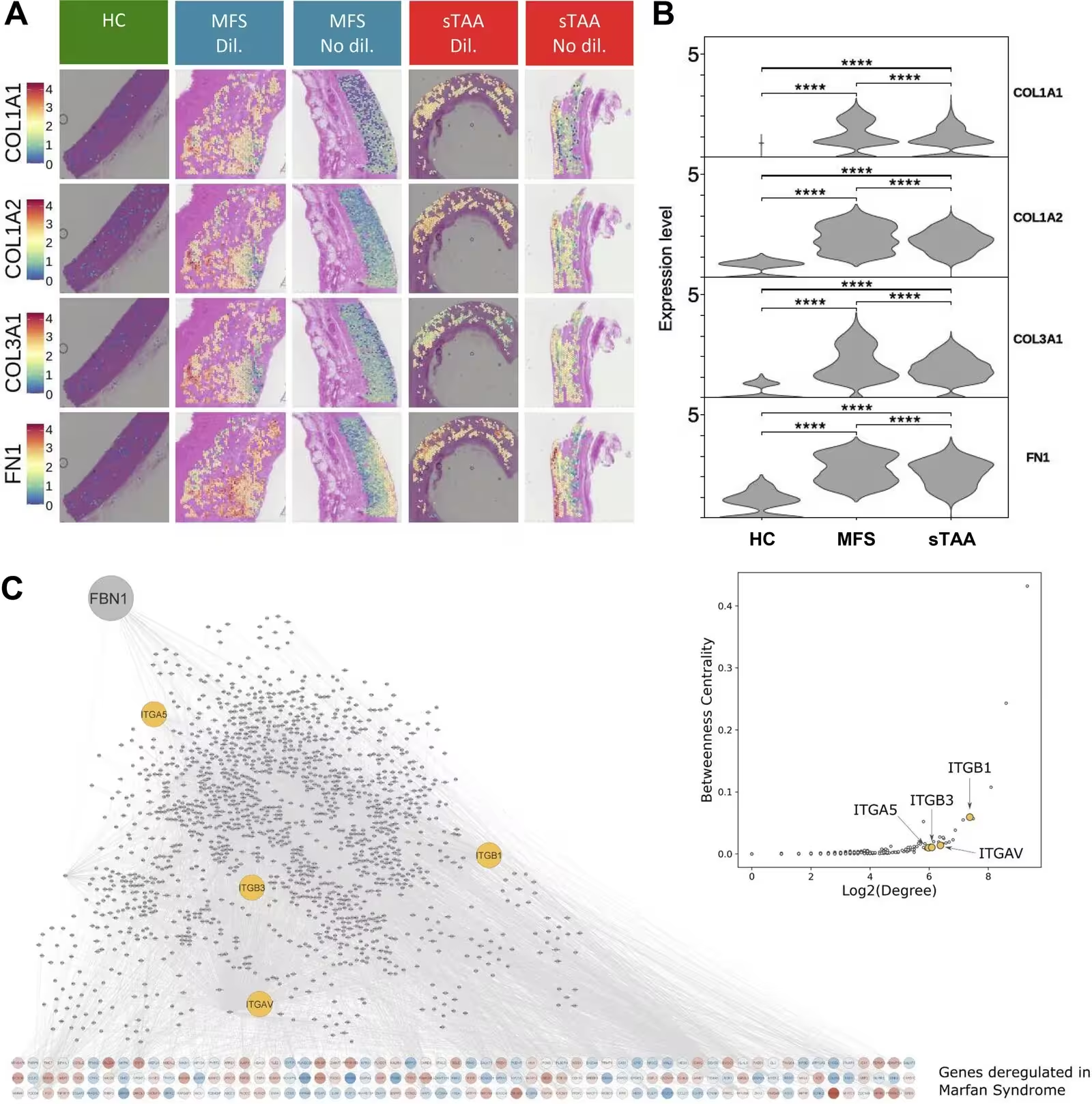
Figure S2: Collagen-related genes are upregulated in MFS and sTAA aortas, and integrins emerge as central nodes in aneurysm pathogenesis . (A and B) Spatial transcriptomics images (A) and relative violin plots (B), depicting the expression levels of genes encoding COL1A1 (first row), COL1A2 (second row), COL3A1 (third row), and fibronectin (FN1, fourth row) on thoracic aortas from HC subjects and patients with MFS or sTAA, both in dilated and non-dilated aneurysmal regions. The violin plots show the expression levels of gene groups in terms of spatial density distribution in cell clusters with more than 45% VSMCs. ****P < 0.0001. (C) Integrins play a key role in the protein-protein interaction (PPI) subnetwork relevant to MFS. This network connects FBN1 to genes deregulated in aortic smooth muscle cells from MFS patients through known PPIs. Genes upregulated in MFS are shown in red; downregulated genes in blue. Integrins (ITGB1, ITGAV, ITGB3, and ITGA5) are highlighted in yellow. The scatterplot
Collagen-related genes are upregulated in MFS and sTAA aortas, and integrins emerge as central nodes in aneurysm pathogenesis . (A and B) Spatial transcriptomics images (A) and relative violin plots (B), depicting the expression levels of genes encoding COL1A1 (first row), COL1A2 (second row), COL3A1 (third row), and fibronectin (FN1, fourth row) on thoracic aortas from HC subjects and patients with MFS or sTAA, both in dilated and non-dilated aneurysmal regions. The violin plots show the expression levels of gene groups in terms of spatial density distribution in cell clusters with more than 45% VSMCs. ****P < 0.0001. (C) Integrins play a key role in the protein-protein interaction (PPI) subnetwork relevant to MFS. This network connects FBN1 to genes deregulated in aortic smooth muscle cells from MFS patients through known PPIs. Genes upregulated in MFS are shown in red; downregulated genes in blue. Integrins (ITGB1, ITGAV, ITGB3, and ITGA5) are highlighted in yellow. The scatterplot Credit: Sara Rega et al. / The Journal of experimental medicine

Halting Aortic Growth in Preclinical Animal Models

To determine whether these cellular benefits translated into living organisms, the researchers tested both drugs in two distinct mouse models representing genetic Marfan syndrome and chemically induced sporadic aneurysms. Animals treated with vehicle solutions showed progressive, severe dilation of both the ventriculo-aortic annulus and the Valsalva sinus over several weeks.

Echocardiographic monitoring revealed that administering Cilengitide three times a week effectively limited aortic dilation at both anatomical sites in both murine models. By contrast, animals treated with the broad-spectrum inhibitor GLPG0187 showed no statistical difference in aortic diameter compared to untreated diseased controls. Histological staining with Masson trichrome further confirmed that targeted inhibition successfully reduced abnormal collagen deposition and fibrosis within the medial layer of the aortic wall.
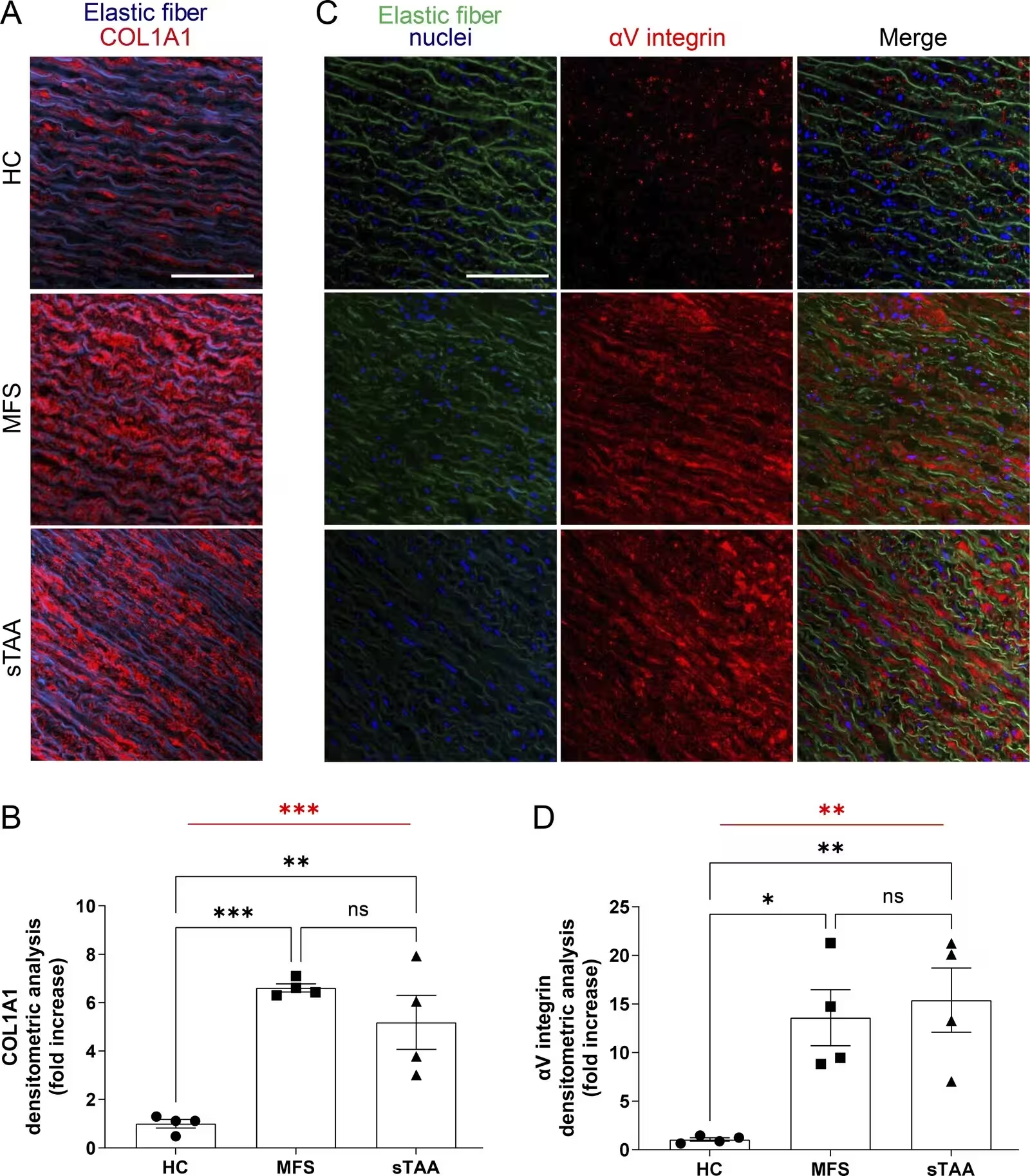
Figure 2: COL1A1 and αV integrin are more abundant in tunica media of MFS-TAA and sTAA than in HC patients. (A and B) Representative immunofluorescence images (A) and quantification (B) of COL1A1 expression in the tunica media of HC, MFS-TAA, and sTAA aortas. (C and D) Representative immunofluorescence images (C) and quantification (D) of αV integrin expression in the tunica media of HC aortas, MFS-TAA, and sTAA. Nuclei are stained with DRAQ5 and shown in blue. In all immunofluorescence images, the tunica intima and adventitia are not visible and are localized in the upper right and lower left corners, respectively. Scale bar = 100 μm. Data are shown as mean ± SEM, n = 4/group. One-way ANOVA (in red): **P < 0.01, ***P < 0.001. Tukey’s post hoc test (in black): *P < 0.05, **P < 0.01, and ***P < 0.001. Experimental data were verified in at least two independent experiments.
COL1A1 and αV integrin are more abundant in tunica media of MFS-TAA and sTAA than in HC patients. (A and B) Representative immunofluorescence images (A) and quantification (B) of COL1A1 expression in the tunica media of HC, MFS-TAA, and sTAA aortas. (C and D) Representative immunofluorescence images (C) and quantification (D) of αV integrin expression in the tunica media of HC aortas, MFS-TAA, and sTAA. Nuclei are stained with DRAQ5 and shown in blue. In all immunofluorescence images, the tunica intima and adventitia are not visible and are localized in the upper right and lower left corners, respectively. Scale bar = 100 μm. Data are shown as mean ± SEM, n = 4/group. One-way ANOVA (in red): **P < 0.01, ***P < 0.001. Tukey’s post hoc test (in black): *P < 0.05, **P < 0.01, and ***P < 0.001. Experimental data were verified in at least two independent experiments. Credit: Sara Rega et al. / The Journal of experimental medicine

Potential Pathways Toward Clinical Translation

The study authors emphasize that because Cilengitide has already undergone extensive human testing and pharmacokinetic profiling in oncology clinical trials, its potential repurposing for cardiovascular conditions could move forward efficiently. While broader systemic blockade of transforming growth factor-beta often causes severe side effects because of its diverse physiological roles throughout the body, targeting specific integrin receptors offers a way to selectively suppress local fibrosis.

Future clinical investigations will need to evaluate whether Cilengitide should be administered as a standalone therapy or combined with standard medications such as beta-blockers or angiotensin receptor blockers. If successful in upcoming human trials, this precision pharmacological approach could finally provide an etiological treatment option for patients facing the constant risk of aortic dissection and rupture.

The research was published in The Journal of experimental medicine on December 7, 2026.

Fact Checked

This article has been fact checked for accuracy, with information verified against reputable sources. Learn more about us and our editorial process.

Last reviewed on .

Article history

  • Latest version

Reference(s)

  1. Rega, Sara., et al. “Cilengitide limits syndromic and sporadic thoracic aortic aneurysm by targeting specific αV integrin.” Journal of Experimental Medicine, vol. 223, no. 12, November 11, 2026 Rockefeller University Press, doi: 10.1084/jem.20260700. <https://doi.org/10.1084/jem.20260700>.

Cite this page:

Kumar, Rohan. “Targeted Drug Halts Dangerous Aortic Aneurysm Growth in Preclinical Study.” BioScience. BioScience ISSN 2521-5760, 12 November 2026. <https://www.bioscience.com.pk/en/subject/biotechnology/cilengitide-limits-syndromic-and-sporadic-thoracic-aortic-aneurysm-by-targeting-specific-av-integrin>. Kumar, R. (2026, November 12). “Targeted Drug Halts Dangerous Aortic Aneurysm Growth in Preclinical Study.” BioScience. ISSN 2521-5760. Retrieved November 12, 2026 from https://www.bioscience.com.pk/en/subject/biotechnology/cilengitide-limits-syndromic-and-sporadic-thoracic-aortic-aneurysm-by-targeting-specific-av-integrin Kumar, Rohan. “Targeted Drug Halts Dangerous Aortic Aneurysm Growth in Preclinical Study.” BioScience. ISSN 2521-5760. https://www.bioscience.com.pk/en/subject/biotechnology/cilengitide-limits-syndromic-and-sporadic-thoracic-aortic-aneurysm-by-targeting-specific-av-integrin (accessed November 12, 2026).
End of the article