Clam Blood Nano-Vesicles Reveal Hidden Metabolic Signals
Researchers isolate delicate marine biological shuttles and prove the feasibility of tracking aquatic ecosystem health through water sampling.
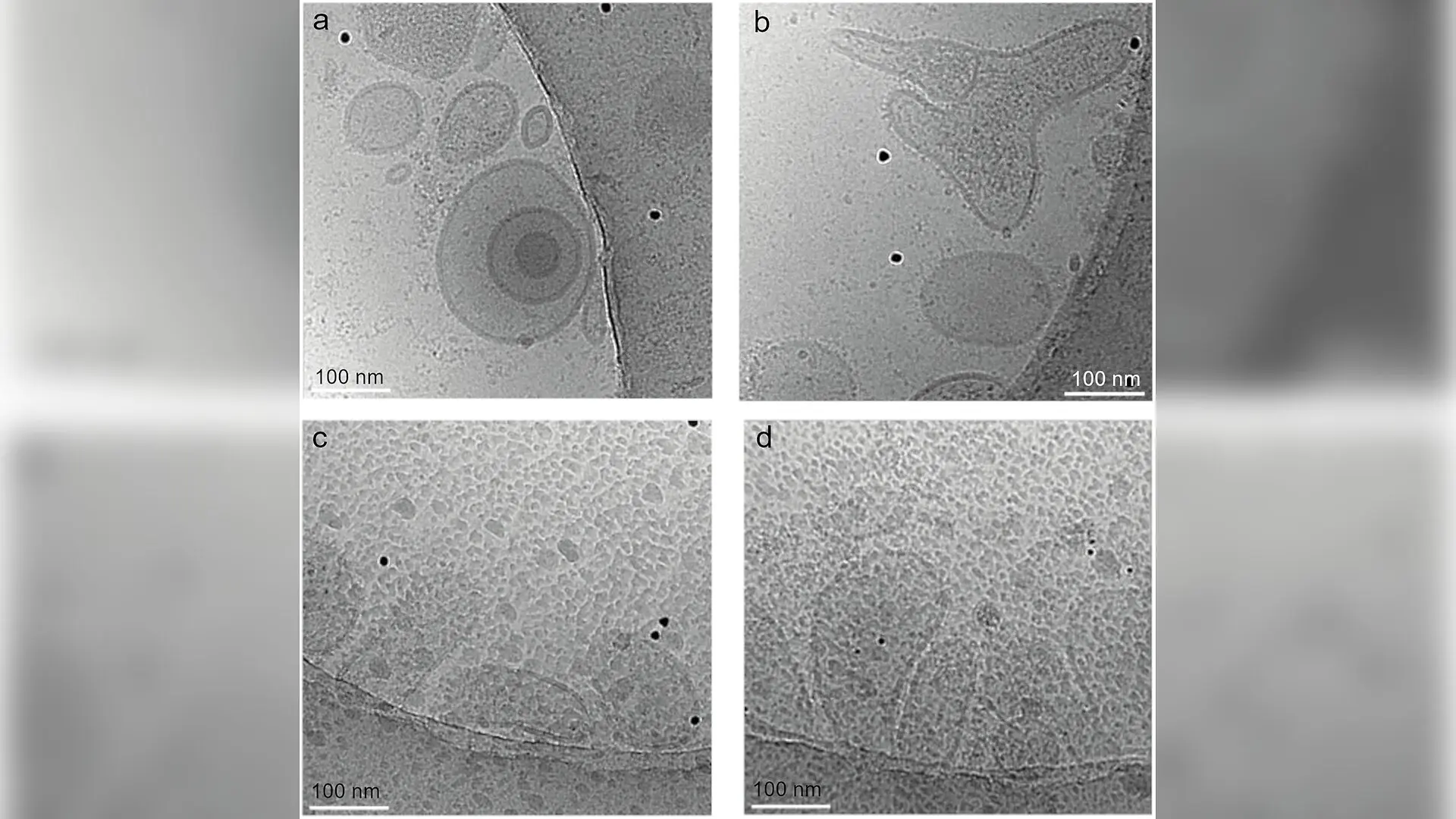
Manila clams release tiny membrane-enclosed packages into their bodily fluids and surrounding water that could soon transform how scientists monitor marine ecosystems and climate stress. These microscopic parcels, known as extracellular vesicles, act as intercellular shuttles carrying proteins, lipids, and metabolites between cells. While researchers have studied these nano-vesicles extensively in humans and mammalian models, their presence and functions in marine invertebrates have remained largely unexplored until now. A team of international researchers has developed and optimized reliable laboratory protocols to isolate and analyze these elusive biological structures from the hemolymph and surrounding aquatic environments of the widely farmed bivalve species, published in PLOS One.
Overcoming Technical Hurdles in Marine Invertebrate Biology
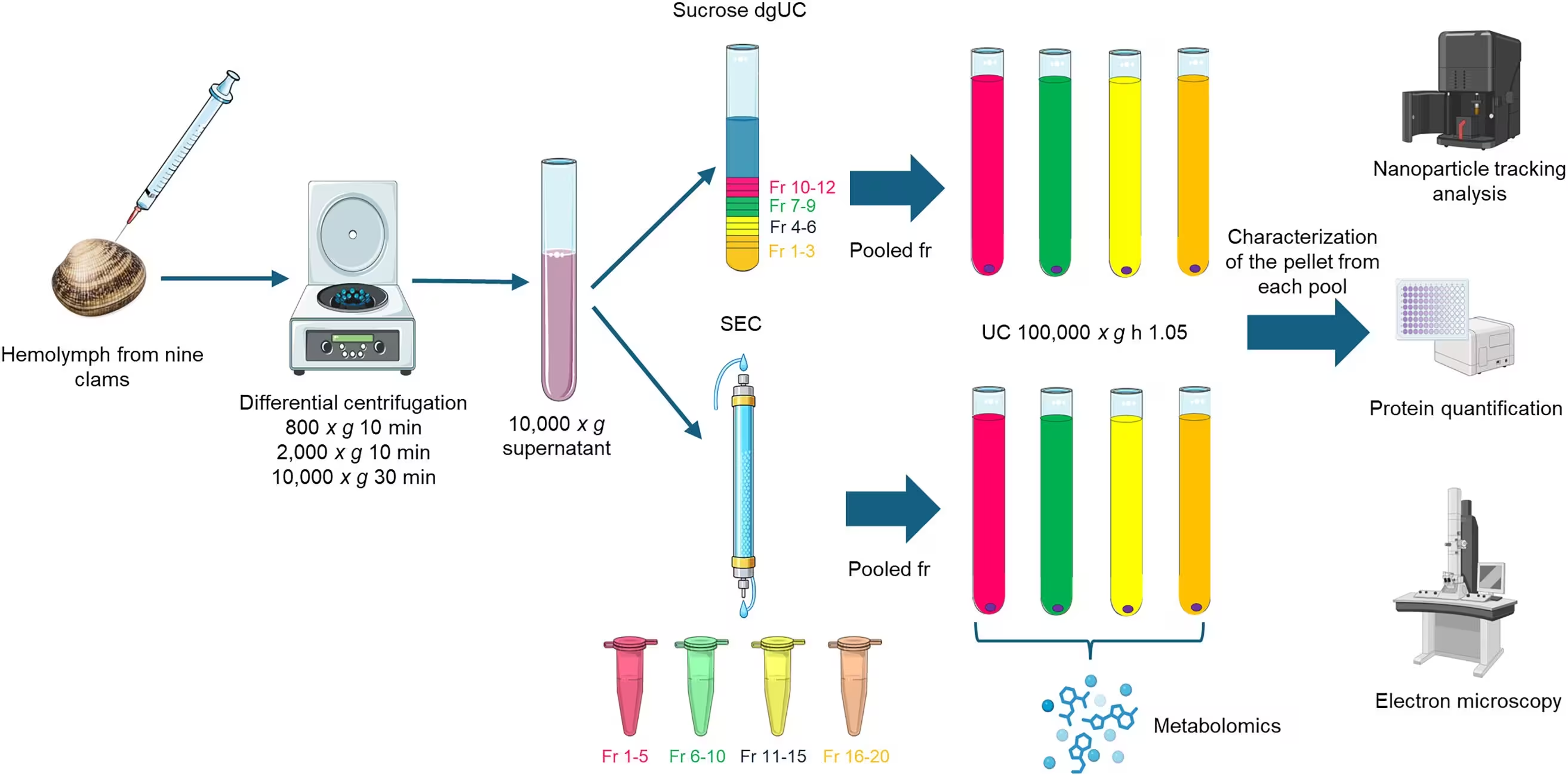
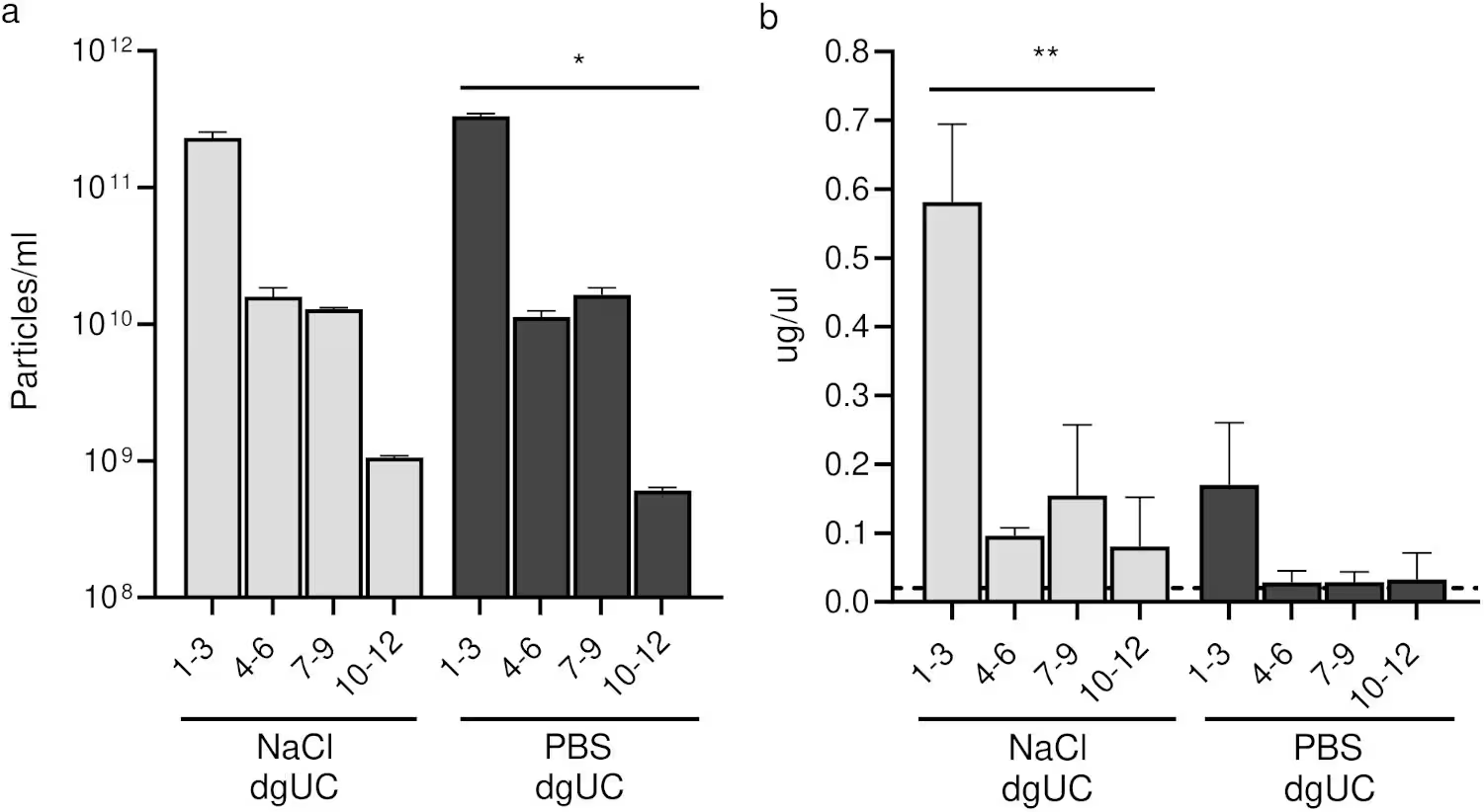
Extracting pure extracellular vesicles from marine bivalves presents unique challenges due to the complex chemical makeup of invertebrate hemolymph, which is the circulatory fluid roughly equivalent to blood. Standard laboratory buffers often trigger unwanted salt precipitates that interfere with downstream analysis. To solve this problem, the authors compared standard phosphate-buffered saline against an isosmotic diluent made from deionized water supplemented with 3.2 percent sodium chloride. Testing both sucrose density gradient ultracentrifugation and size exclusion chromatography revealed that while ultracentrifugation yielded cleaner vesicle preparations, chromatography successfully recovered higher nanoparticle concentrations necessary for detailed molecular profiling.
Using nanoparticle tracking analysis alongside transmission electron microscopy and cryo-electron microscopy, the team confirmed that the isolated vesicles predominantly measured under 200 nanometers in diameter. Cryo-electron microscopy further demonstrated that utilizing the sodium chloride-supplemented diluent prevented the formation of salt aggregates that otherwise degraded image quality in phosphate-buffered saline samples. However, standard mammalian antibodies directed against common vesicle markers such as CD9, TSG101, and integrin-beta failed to show cross-reactivity with clam proteins, highlighting the evolutionary divergence between humans and bivalves and pointing to the urgent need for species-specific molecular tools.
Unlocking Unique Metabolic Profiles Without Fully Annotated Genomes
Because the Manila clam genome lacks complete functional annotation, traditional proteomic and transcriptomic tracking methods remain difficult. To circumvent this barrier, the investigators turned to liquid chromatography-mass spectrometry to map the metabolic cargo contained within the isolated vesicles. Their analysis detected between 62 and 81 distinct metabolites across the tested fractions, revealing that the vesicle-enriched chromatography fractions possessed a distinct chemical signature compared to the whole hemolymph supernatant.
Crucially, several vital metabolites, including adenosine monophosphate, oxidized glutathione, nicotinamide adenine dinucleotide, and key glycolytic intermediates, appeared exclusively within the vesicle-enriched fractions. These specific compounds play central roles in cellular energy metabolism and redox regulation. The findings suggest that Manila clam vesicles actively package and transport specific metabolic regulators, opening up new avenues for assessing physiological health in aquatic species through metabolomic screening.
Detecting Biological Nano-Vesicles in Aquatic Environments
To evaluate whether clams actively shed these vesicles into their natural habitat, the researchers conducted a pilot study holding groups of five or ten Manila clams in controlled water volumes for 48 hours. Using tangential flow filtration followed by ultrafiltration, they concentrated the particulate matter and compared the samples against blank water controls and control bottles containing sterilized empty shells. Nanoparticle tracking analysis, atomic force microscopy, and a specialized gold nanoparticle aggregation assay confirmed the presence of genuine membranous, vesicle-like structures alongside mineral particles shed from the calcium carbonate shells.
Although the presence of background microorganisms and shell-associated particles means that some vesicles likely originated from co-existing life forms, the work demonstrates the fundamental feasibility of recovering animal-derived vesicles from water. The authors suggest that this non-invasive sampling approach could eventually form the basis of advanced environmental biomonitoring programs. By tracking changes in vesicle abundance and cargo composition, conservationists and aquaculture managers might soon gain a powerful tool to evaluate how climate change, heat waves, and pollution impact marine ecosystems.
The research was published in PLOS One on November 17, 2026.
This article has been fact checked for accuracy, with information verified against reputable sources. Learn more about us and our editorial process.
Last reviewed on .
Article history
- Latest version
Reference(s)
- Moccia, Valentina., et al. “Tailored isolation and metabolomic profiling of extracellular vesicles in Manila clam (Ruditapes philippinarum) hemolymph and insights into water-derived vesicles.” PLOS One, vol. 21, no. 11, November 17, 2026, pp. e0362957 Public Library of Science (PLoS), doi: 10.1371/journal.pone.0362957. <https://doi.org/10.1371/journal.pone.0362957>.
Cite this page:
- Posted by Hassan Raza