300-Million-Year-Old Fish Brain Preserved in Coal: CT Scan Reveals Ancient Neural Architecture
Scientists examine an exceptionally preserved fossil to trace vertebrate brain evolution across hundreds of millions of years.
Researchers have uncovered a coal‑bearing fossil from northwest England that contains a remarkably intact brain dating back roughly 300 million years. The find offers an unprecedented window into the neuroanatomy of one of the earliest ray‑finned fishes and may force a rethink of how vertebrate brain evolution is studied. The results, appearing in the Proceedings of the National Academy of Sciences, suggest that fossilized skulls can retain far more soft‑tissue detail than previously assumed, opening new avenues for reconstructing the neural architecture of long‑lost species.
Rare Brain Preservation Sheds Light on Early Fish Morphology
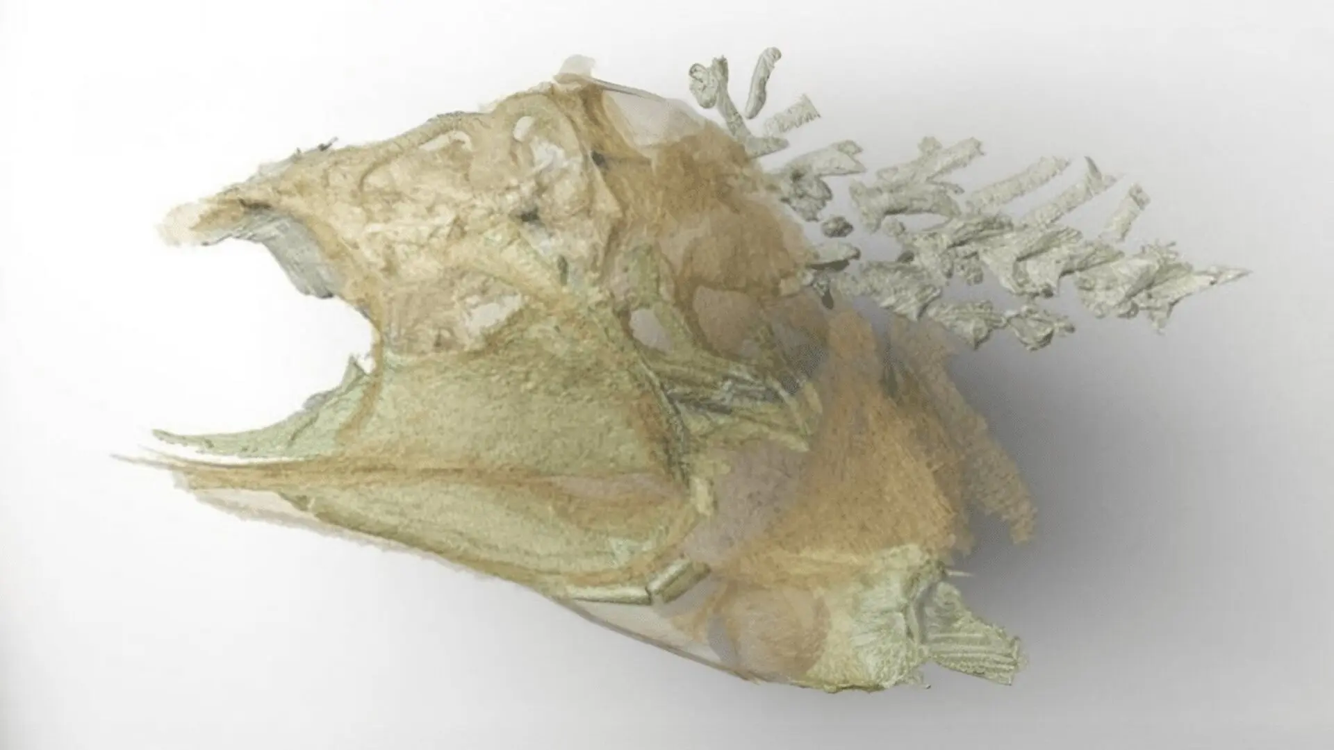
Trawdenia planti, a diminutive ray‑finned fish that swam in swampy habitats covering present‑day Lancashire during the Carboniferous, is the source of the specimen. After death the animal sank into fine sediment trapped between coal seams, where unique conditions halted decomposition. While bone, scale and tooth fragments often survive, soft tissues usually decay within days; neural tissue is especially vulnerable, making the preservation of a brain an extraordinary rarity.
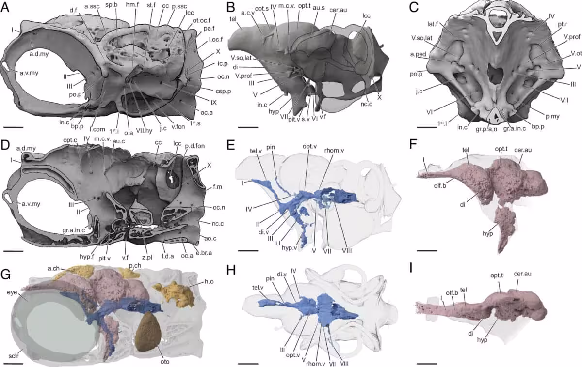
By employing high‑resolution CT scanning and digital modeling, the team identified remnants of the brain, its surrounding membranes, and ventricular chambers that once held cerebrospinal fluid. The data produced a detailed three‑dimensional reconstruction of the brain’s position inside the skull, representing one of the most complete fossilized neural structures ever recorded. Dr. Abigail Caron highlighted the significance, noting,
“Soft tissue preservation, in general, is not common in the fossil record, and usually what gets preserved are things like skin or muscles. It’s quite rare for neural tissues to be preserved at all because they decay so quickly.” She added, “So, the importance of this specimen is that we can now study brain evolution in similar fossils where we only have the bony parts or the infill.”
New Pathways for Reconstructing Ancient Vertebrate Brains
The PNAS paper (https://www.pnas.org/doi/10.1073/pnas.2610438123) argues that the brain of Trawdenia planti occupied nearly the entire cranial cavity, a pattern that diverges from many fossil vertebrates where the brain fills only a fraction of the skull. If early ray‑finned fishes shared this tight brain‑skull coupling, paleontologists could infer brain morphology simply from well‑preserved braincases.
Because skulls survive far more often than soft tissues, this approach could dramatically expand the pool of specimens available for neuro‑evolutionary research. Existing museum collections might thus become a trove of neurological data without the need for additional rare finds. As imaging resolution continues to improve, researchers anticipate uncovering further hidden anatomical clues within long‑stored fossils.
A Small Carboniferous Fish Sheds Light on a Major Evolutionary Question
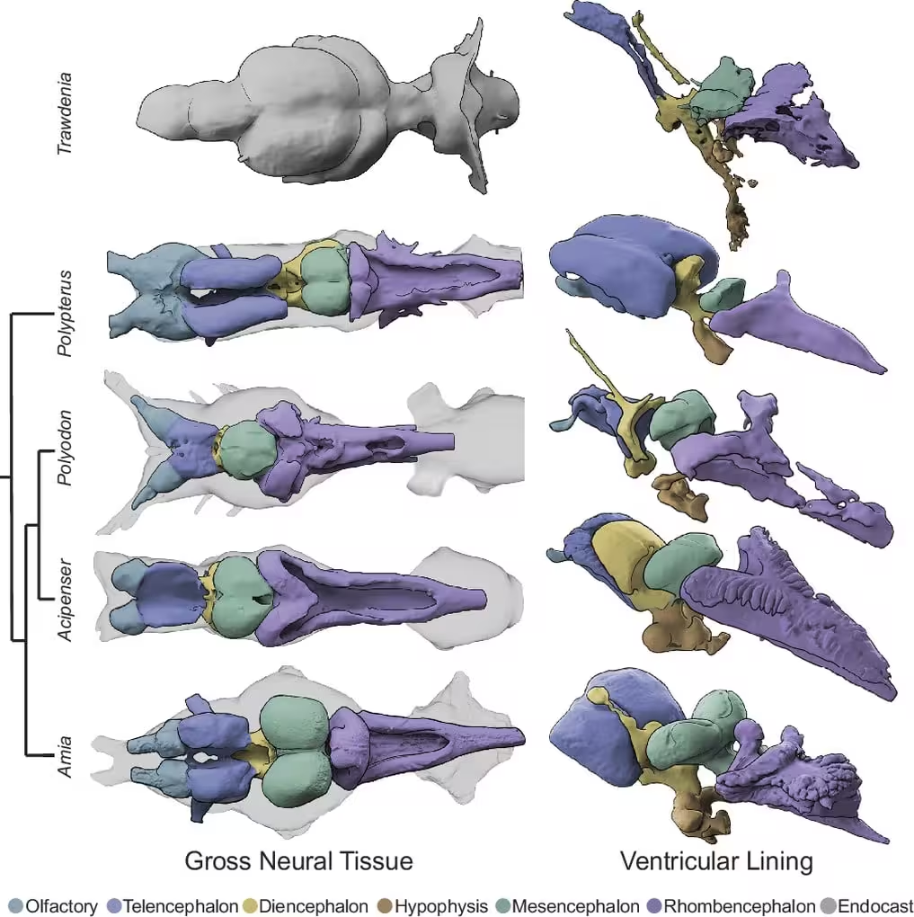
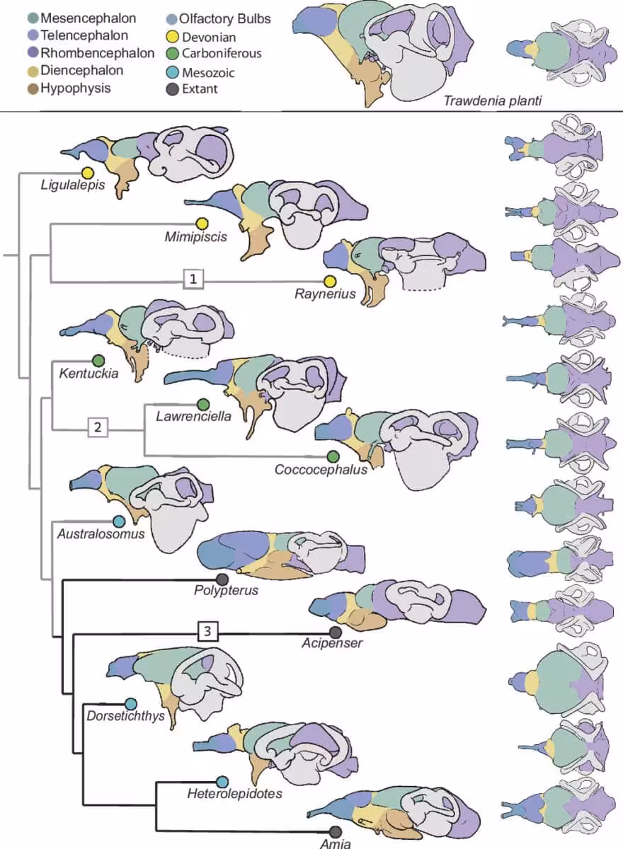
Ray‑finned fishes dominate modern aquatic life, representing nearly 99 percent of living fish species and about half of all vertebrates. Yet their deep evolutionary history remains murky, as early members display a blend of primitive and derived traits. Dr. Michael Coates likens the base of the ray‑finned tree to “a bush at the bottom of the evolutionary tree.” The preserved brain of Trawdenia planti provides a rare data point to untangle these relationships. Its internal layout shares features with contemporary paddlefish and sturgeons, such as a cerebellum that wraps around the central brain region. Dr. Coates explains,
“What we’re learning by looking at the shapes of the soft tissues is that it’s not their relative sizes that matter; it’s how they’re packed together inside the skull.” He added, “We might be seeing the earliest radiation of fishes that nowadays are represented by paddlefish and sturgeons.”
These findings imply that brain organization may reveal evolutionary signals that skeletal anatomy alone cannot capture.
High‑Resolution Scanning Revives a Century‑Old Coal Fossil
The specimen has been part of scientific collections since its discovery by Lancashire coal miners in 1888. The fossil was split into two halves that entered the Natural History Museum in London separately, and for many decades researchers examined only the skeletal elements because earlier techniques could not detect soft‑tissue remnants.
Advances in micro‑CT and image‑processing now allow scientists to peer inside the rock without harming the specimen. Dr. Caron remarks,
“It’s possible that we just didn’t have the technology before to look for that kind of signature, but this kind of preservation also would only happen in very special circumstances.” She also explained, “There are definitely a lot more specimens out there that have reasonably good brain case morphology than there are specimens with good soft tissue preservation.” Finally, she concluded, “So, that really expands the data set that you can use to study brain evolution across these different fossils.”
The case demonstrates how contemporary imaging methods can turn long‑known museum pieces into fresh sources of scientific insight, revealing details that have remained hidden for generations.
This article has been fact checked for accuracy, with information verified against reputable sources. Learn more about us and our editorial process.
Last reviewed on .
Article history
- Latest version
Reference(s)
- Caron, Abigail M.., et al. “Neural tissues and chondrostean traits in a Carboniferous actinopterygian.” Proceedings of the National Academy of Sciences, vol. 123, no. 27, June 29, 2026 National Academy of Sciences, doi: 10.1073/pnas.2610438123. <https://www.pnas.org/doi/10.1073/pnas.2610438123>.
Cite this page:
- Posted by Hassan Raza