Ancient One-Eyed Sea Worm May Have Seeded Human Vision and the Pineal Gland
A 600‑million‑year‑old one‑eyed sea creature may hold the key to human vision, revealing an unexpectedly strange evolutionary twist.
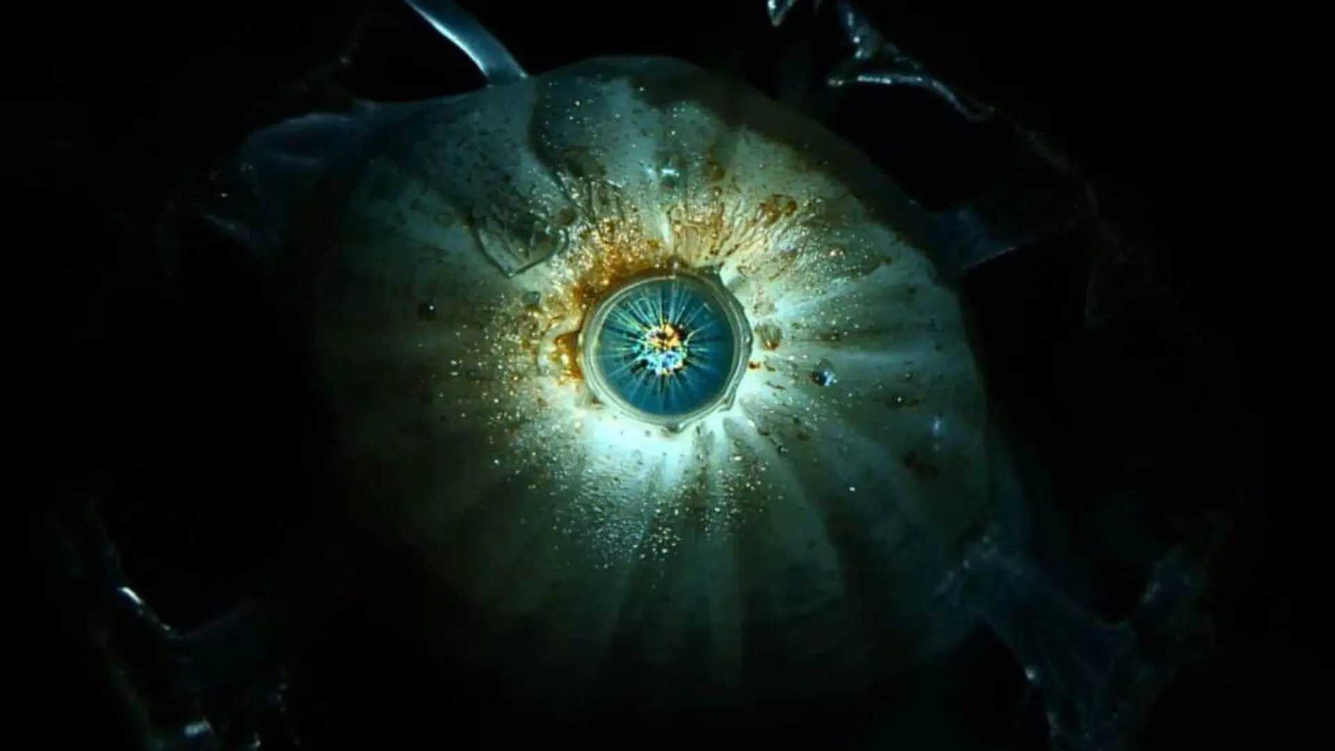
New research suggests that a tiny, worm‑shaped marine animal that thrived roughly 600 million years ago may have housed the earliest blueprint for the visual system that eventually gave rise to human eyes. While this filter‑feeding creature abandoned the paired eyes typical of its relatives, it preserved a cluster of light‑sensitive cells at the center of its head, forming a rudimentary median eye.
Scientists from Lund University and the University of Sussex argue that portions of this primitive organ were later co‑opted into the paired eyes of vertebrates. Their findings, detailed in Current Biology, also link the lingering central eye structure to the pineal gland, the brain region that governs circadian rhythms.
From Filter Feeder to Vision Pioneer
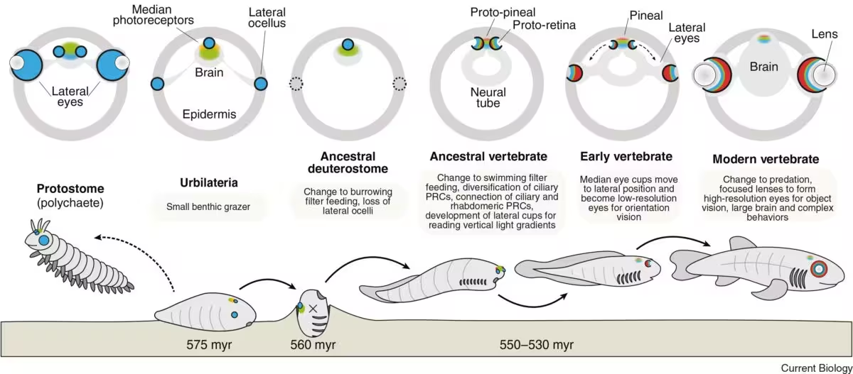
The organism at the heart of the study was a sedentary, plankton‑filtering worm‑like animal. Earlier members of its lineage possessed some form of paired eyes, though the precise capabilities of those eyes remain uncertain.
“We cannot tell whether the paired eyes in this branch were merely light‑sensitive patches or capable of forming images,” said Dan‑E Nilsson, professor emeritus of sensory biology at Lund University, in a university press release. As the animal’s lifestyle shifted toward a more passive filter‑feeding mode, the selective pressure to maintain complex vision waned, leading to the loss of the lateral eyes while retaining a central photoreceptive patch.
The residual median eye could not resolve detailed images but was sufficient to detect light‑dark transitions, track day‑night cycles, and provide a rudimentary sense of orientation. Over evolutionary time, descendants that reverted to an active, swimming lifestyle would have found image‑forming vision advantageous once again.
Recycling a Central Sensor for Paired Vision
When the lineage re‑acquired a motile existence, the retained central photoreceptive system may have been repurposed to form new lateral eyes. Rather than constructing a visual apparatus from scratch, evolution appears to have remodeled existing cells and neural pathways, integrating them into the emerging paired eye structures.

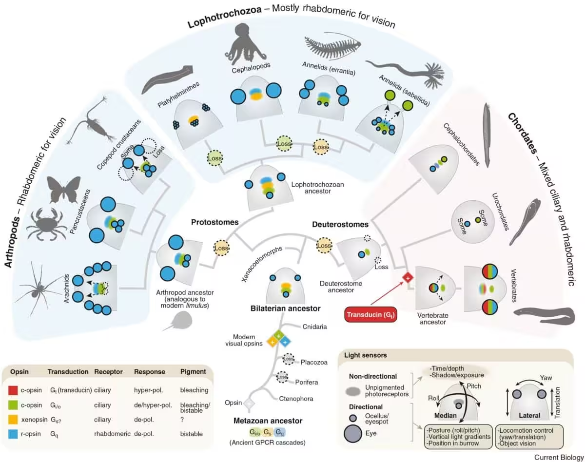
This scenario clarifies why vertebrate retinas originate from brain tissue, unlike the lateral eyes of insects and cephalopods that develop from surface ectoderm. The ancestral median organ’s association with central nervous tissue would have preserved that developmental link when its components were incorporated into paired eyes.
In vertebrates, the retina—a layered network of photoreceptors and interneurons at the back of the eye—processes incoming light before transmitting signals deeper into the brain. The new model traces a plausible evolutionary trajectory from a simple central light sensor to the sophisticated paired retinas observed in modern vertebrates.
Why Vertebrate Eyes Diverge from Insect and Cephalopod Vision
Most bilaterally symmetric animals develop eyes on the sides of the head, yet vertebrate retinas emerge as extensions of the brain. Nilsson explains that the loss of the original paired eyes in the vertebrate lineage forced the remaining central organ onto a distinct developmental pathway. When paired vision later reappeared, it did so using elements derived from that central system.
The authors stress that this does not imply superiority of one eye design over another; insects, cephalopods and vertebrates each arrived at functional visual systems through separate evolutionary routes. The focus of the study is the specific developmental relationship that ties vertebrate retinas to the brain.
By mapping the distribution and characteristics of photoreceptor cells across diverse animal groups, the team identified patterns that preserve hidden evolutionary signals, even when external morphology no longer reflects those origins.
A Legacy Inside the Brain: The Pineal Gland
Not all components of the ancient median eye were incorporated into paired retinas. The researchers propose that the pineal gland—a small endocrine organ within the vertebrate brain that secretes melatonin—represents a surviving fragment of the original central eye.
Melatonin production is tightly linked to the circadian rhythm, the roughly 24‑hour cycle that orchestrates sleep‑wake patterns. Because light is a primary cue for resetting this internal clock, the pineal gland’s light‑sensitive ancestry fits the hypothesis that it evolved from an ancient photoreceptive structure used to monitor day and night.
This article has been fact checked for accuracy, with information verified against reputable sources. Learn more about us and our editorial process.
Last reviewed on .
Article history
- Latest version
Reference(s)
- <https://www.cell.com/current-biology/fulltext/S0960-9822(25)01676-8>.
- “One‑eyed creature gave rise to our modern eyes.”, July 15, 2026 <https://www.lunduniversity.lu.se/article/one-eyed-creature-gave-rise-our-modern-eyes>.
Cite this page:
- Posted by Hassan Raza