Hidden Knee Swelling Revealed in At-Risk Patients Over 12 Months
High-resolution imaging exposes continuous fluid shifts inside the joint that standard medical scans completely miss.
Knee joints at risk of osteoarthritis undergo hidden fluid shifts over the course of a year, even when standard medical scans show no visible change in joint inflammation. Researchers examining data from 680 knees found that while traditional categorical grading missed subtle fluctuations, precise volumetric measurements revealed continuous fluid dynamics. The work was published in PloS one by a multi-institutional team analyzing the Osteoarthritis Initiative.
Osteoarthritis has long been viewed as a simple wear-and-tear condition of the cartilage, but modern research highlights low-grade joint inflammation, known as effusion-synovitis, as a major driver of pain and structural decline. To understand what fuels this internal swelling over time, the investigators tracked participants from an enriched incidence cohort at high risk for developing knee osteoarthritis. The average participant age was 60.2 years, and 66.9 percent of the knees belonged to female participants.
The team quantified synovial fluid volume using magnetic resonance imaging at the start of the study and again 12 months later. They also measured baseline clinical factors, metabolic markers like diabetes and blood pressure, and physical performance tests including walking speed and quadriceps muscle strength.
Why Standard Medical Scans Miss Subtle Joint Changes
When the researchers evaluated the scans using traditional semi-quantitative scoring systems, more than 75 percent of the knees showed no grade shift over the 12-month period. However, detailed three-dimensional volumetric segmentation exposed considerable underlying instability. Even among knees classified as completely stable by standard grading, the average absolute volume change was 2.10 milliliters.
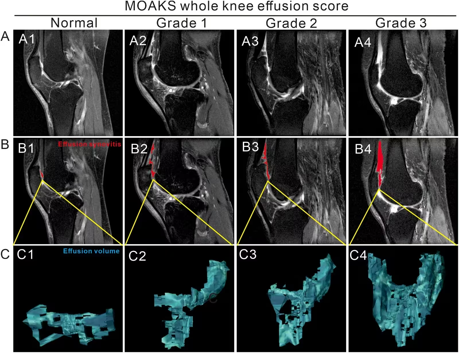
Across the entire cohort of 680 knees, 45.0 percent experienced volumetric expansion greater than 0.5 milliliters, while 37.8 percent showed spontaneous regression dropping by more than 0.5 milliliters. Another 17.2 percent remained stable within a half-milliliter margin. This high-resolution approach captured subtle inflammatory trajectories that conventional floor and ceiling thresholds completely obscure.
What Drives Sustained Inflammation Inside the Knee?
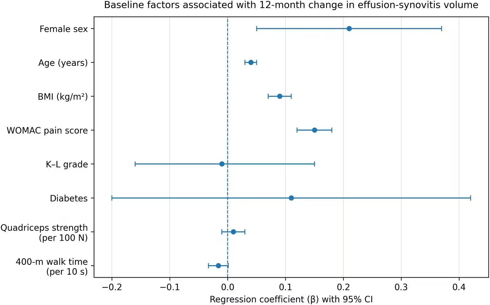
Statistical models revealed that specific baseline characteristics were independently linked to greater increases in joint fluid volume over the 12-month window. Female sex, older age, higher body mass index, and higher Western Ontario and McMaster Universities Arthritis Index pain scores were all significantly associated with worsening inflammation. Specifically, higher body mass index showed a regression coefficient of 0.09, and baseline pain scores tracked closely with subsequent fluid accumulation.
In contrast, structural severity assessed via Kellgren-Lawrence radiographic grades showed no significant association with longitudinal changes in fluid volume. Diabetes status, quadriceps strength, and 400-meter walk times also failed to show significant overall links to 12-month inflammation shifts. The data point to a relative dissociation between structural damage seen on standard X-rays and the active inflammatory processes occurring within the joint synovium.
What These Findings Mean for Future Patient Care
The study authors note that evaluating quantitative changes over a full 12-month window captures sustained intra-articular inflammatory drive rather than temporary day-to-day fluid fluctuations caused by acute physical stress. Factors such as elevated body mass index and persistent pain appear to act as key determinants in knees that resist spontaneous inflammatory reduction.
Several limitations should be considered when interpreting these results. The observational design prevents definitive causal claims, and unmeasured lifestyle factors or occupational loading could introduce residual confounding. Furthermore, the 12-month follow-up period limits long-term extrapolation, and comprehensive metabolic profiling beyond basic blood pressure and diabetes status was unavailable.
Despite these constraints, the work suggests that patient-reported pain and demographic risk factors provide vital clues about underlying synovial activity that standard structural imaging misses. Integrating symptom evaluation with advanced volumetric imaging could ultimately improve risk stratification and monitoring for individuals vulnerable to rapid knee osteoarthritis progression.
The research was published in PloS one on November 16, 2026.
This article has been fact checked for accuracy, with information verified against reputable sources. Learn more about us and our editorial process.
Last reviewed on .
Article history
- Latest version
Reference(s)
- Wang, Yang., et al. “Functional performance and 12-month change in effusion-synovitis in knees at risk of osteoarthritis: A longitudinal analysis using the osteoarthritis initiative.” PLOS One, vol. 21, no. 11, November 16, 2026, pp. e0362768 Public Library of Science (PLoS), doi: 10.1371/journal.pone.0362768. <https://doi.org/10.1371/journal.pone.0362768>.
Cite this page:
- Posted by Hassan Raza