Brain Cells Found to Use Dual-Feature System for Vision
Biology

Brain Cells Found to Use Dual-Feature System for Vision

Advanced digital twin models reveal that visual cortical neurons are actively silenced by specific non-preferred images rather than just ignoring them.
By Priya Sharma
Published:
Email this Article
Brain Cells Found to Use Dual-Feature System for Vision
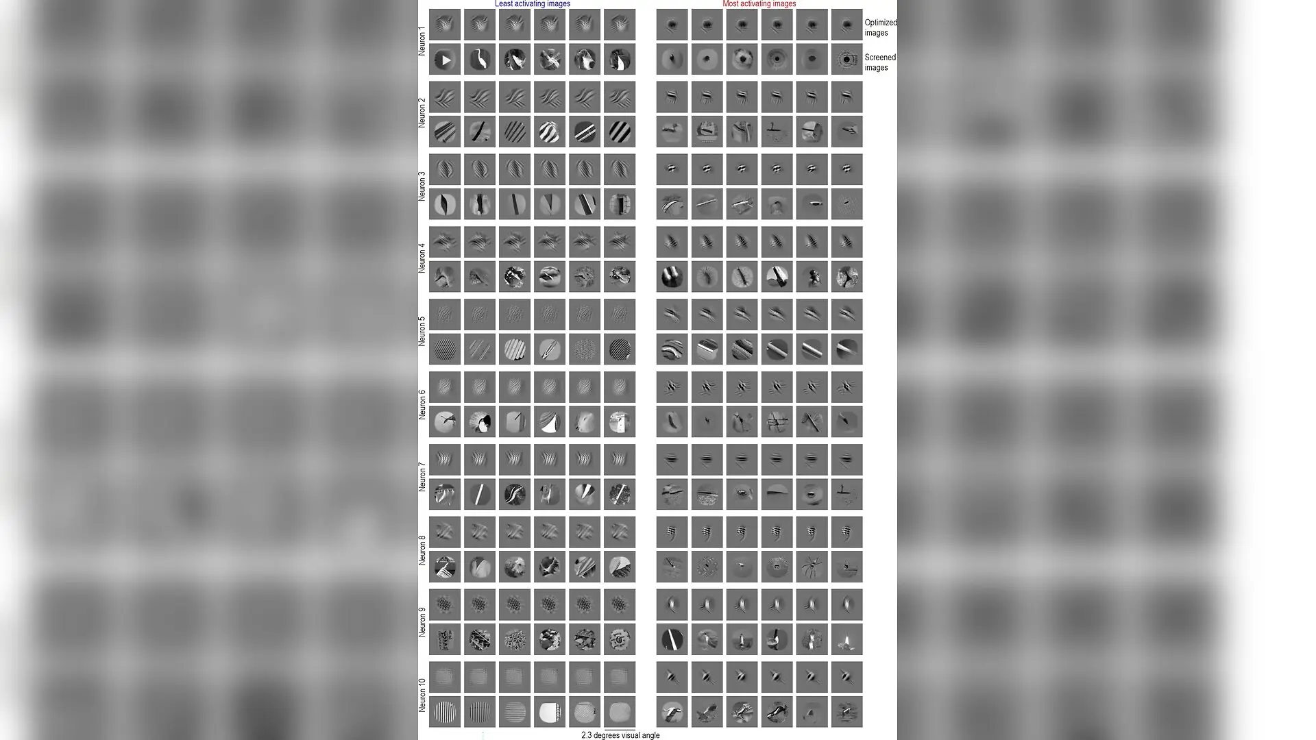
Most activating images (MAIs) and least activating images (LAIs) of example V1 neurons. LAIs (left) and MAIs (right) of 10 example V1 neurons. Per neuron, the top row shows optimized images (least exciting inputs [LEIs] and most exciting inputs [MEIs]), and the bottom row shows screened ImageNet images (LAIs and MAIs). Each image is 2.3 × 2.3 degrees visual angle, with the receptive field of the neuron in the center. Nikos Karantzas et al. / eLife

Brain cells in the visual cortex do much more than simply light up when they spot their favorite picture, as a new study reveals they are actively silenced by completely different images. By combining biological recordings with advanced computer simulations, researchers uncovered a dual-feature coding system operating across both primate and mouse brains.

For decades, neuroscientists viewed individual sensory neurons as dedicated feature detectors. In this classical model, a brain cell remains mostly quiet until a specific stimulus, such as a particular edge orientation or face, enters its receptive field and triggers a surge in electrical firing. Any other visual input is simply ignored.

Yet, real neurons often maintain a steady, non-zero baseline of firing activity when staring at a blank screen. To understand how these cells handle complex natural scenes, a large research team published in eLife deployed deep learning models known as functional digital twins. These artificial intelligence systems learn the exact relationship between visual stimuli and recorded neural responses, allowing scientists to simulate brain activity across millions of images.

How Dual-Feature Selectivity Shapes Firing Rates

The researchers recorded spiking activity from hundreds of neurons in the primary visual area V1 and mid-level area V4 of rhesus macaques, as well as visual areas in mice. When the digital twins screened over one million naturalistic images, they exposed a surprising pattern among non-sparse neurons. These cells did not just fire for preferred features, but they also showed systematic suppression below their baseline when exposed to distinct, non-preferred images.
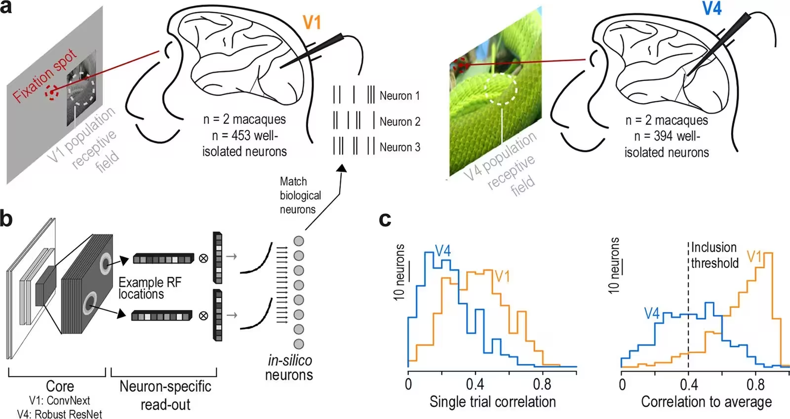
Figure 1: Experimental approach. (a) Experimental recordings from macaque visual areas V1 (453 neurons from two monkeys) and V4 (394 neurons from two monkeys) during presentation of natural images. Monkeys fixated on the screen (fixation spot shown in red). The stimulus was centered on the population receptive field of neurons recorded that day, indicated by the white circle. (b) Architecture of functional ‘digital twin’ models, consisting of a core shared across neurons (V1: first layer of ConvNeXt; V4: layer 3 of Robust ResNet50) and a neuron-specific readout. This design creates in silico neurons that model the response properties of individual biological neurons, with example receptive field locations of two neurons illustrated. (c) Prediction accuracy of the model on test images not used during training, for both V1 (orange, n=75n=75 test images) and V4 (blue, n=150n=150 test images). Left panel shows distribution of single-trial correlations, and right panel displays correlation to the ave
Experimental approach. (a) Experimental recordings from macaque visual areas V1 (453 neurons from two monkeys) and V4 (394 neurons from two monkeys) during presentation of natural images. Monkeys fixated on the screen (fixation spot shown in red). The stimulus was centered on the population receptive field of neurons recorded that day, indicated by the white circle. (b) Architecture of functional ‘digital twin’ models, consisting of a core shared across neurons (V1: first layer of ConvNeXt; V4: layer 3 of Robust ResNet50) and a neuron-specific readout. This design creates in silico neurons that model the response properties of individual biological neurons, with example receptive field locations of two neurons illustrated. (c) Prediction accuracy of the model on test images not used during training, for both V1 (orange, n=75n=75 test images) and V4 (blue, n=150n=150 test images). Left panel shows distribution of single-trial correlations, and right panel displays correlation to the ave Credit: Nikos Karantzas et al. / eLife

Instead of acting like digital on-off switches, these neurons operate on a continuous sliding scale. Their firing rates vary smoothly based on how closely an incoming visual feature matches their preferred excitation anchor versus their suppressive anchor. This bidirectional modulation means a single neuron can continuously encode the contrast between two entirely different visual concepts.

Why Structured Suppression Matters for Vision

Neuroscientists have long known about basic forms of inhibition in the brain, such as cross-orientation inhibition in the primary visual cortex, which helps sharpen tuning curves. However, the new findings demonstrate that suppression is far more complex than previously realized. The least activating images for individual neurons comprised intricate combinations of orientation, spatial frequency, phase, and texture that differed systematically from their excitation preferences.
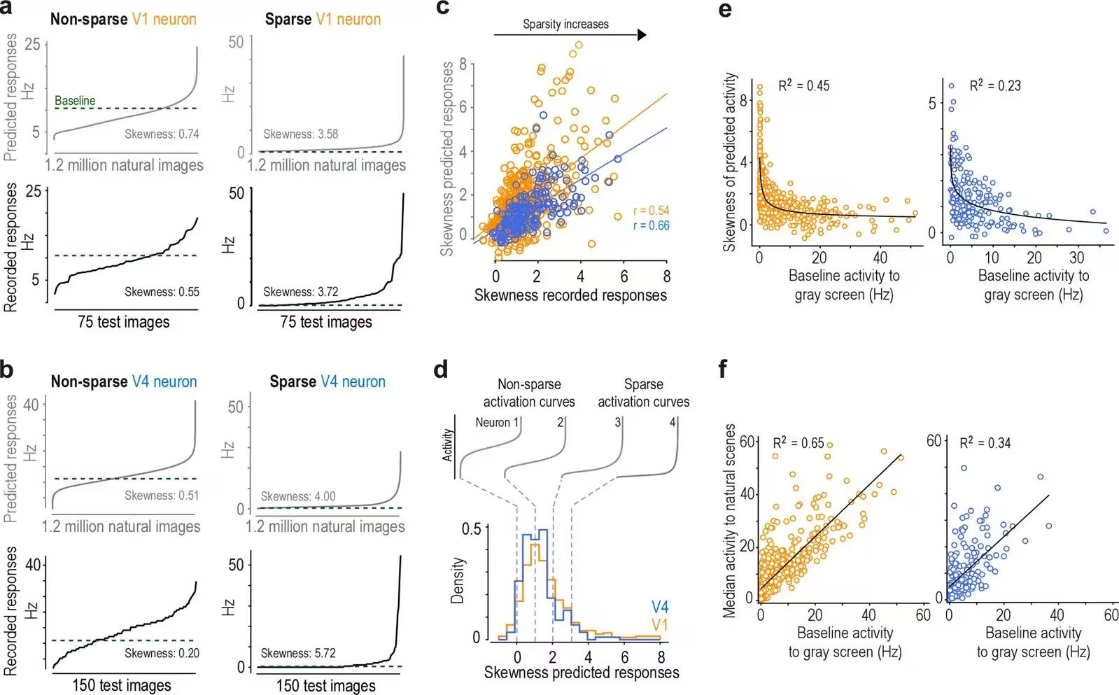
Figure 2: Continuum of single-neuron response sparseness in early and mid-level macaque visual cortex. (a) Response profiles of non-sparse (left) and sparse (right) V1 neurons. Curves display neuronal activity sorted from lowest to highest response, derived from model predictions across 1.2 million ImageNet images (gray, top row) and recorded responses to 75 test images (black, bottom row), averaged over stimulus repeats. We used responses to test images to obtain mean stimulus-driven activity and average out stimulus-unrelated signals. Skewness values quantify lifetime sparsity, with higher values indicating neurons that respond selectively to fewer stimuli while remaining silent to most others. Green dotted lines indicate recorded baseline firing rate (Hz) during gray screen presentation prior to stimulus onset. (b) Comparable response profiles for representative V4 neurons, demonstrating similar variations in lifetime sparsity in this higher-order visual area, with some neurons showing broadl
Continuum of single-neuron response sparseness in early and mid-level macaque visual cortex. (a) Response profiles of non-sparse (left) and sparse (right) V1 neurons. Curves display neuronal activity sorted from lowest to highest response, derived from model predictions across 1.2 million ImageNet images (gray, top row) and recorded responses to 75 test images (black, bottom row), averaged over stimulus repeats. We used responses to test images to obtain mean stimulus-driven activity and average out stimulus-unrelated signals. Skewness values quantify lifetime sparsity, with higher values indicating neurons that respond selectively to fewer stimuli while remaining silent to most others. Green dotted lines indicate recorded baseline firing rate (Hz) during gray screen presentation prior to stimulus onset. (b) Comparable response profiles for representative V4 neurons, demonstrating similar variations in lifetime sparsity in this higher-order visual area, with some neurons showing broadl Credit: Nikos Karantzas et al. / eLife

This structured suppression is conserved across species, appearing in both mice and macaques. By coupling specific excitation with tailored suppression, the brain may significantly increase its representational capacity. Rather than needing a massive population of purely unidirectional cells to encode every possible visual feature, the nervous system uses dual-feature tuning to span a broader dynamic range.

Testing the Limits of Digital Twin Models

Because the core discoveries relied on computer simulations, the team implemented rigorous validation steps to rule out algorithmic artifacts. They tested their findings using independently trained evaluator models with entirely different architectures. These separate neural networks confirmed that the most and least activating stimuli identified by the primary models consistently drove extreme responses in biological neurons.

While the study provides a robust framework for understanding visual coding, several limitations remain. The digital twin models processed cropped images without full contextual surroundings, and the perceptual similarity metrics relied on human rather than macaque judgments. Future research will explore how these opposing excitation and suppression pathways are wired at the microscopic circuit level, shedding light on the fundamental architecture of sensory processing.

The research was published in eLife on November 9, 2026.

Fact Checked

This article has been fact checked for accuracy, with information verified against reputable sources. Learn more about us and our editorial process.

Last reviewed on .

Article history

  • Latest version

Reference(s)

  1. Karantzas, Nikos., et al. “Dual-feature selectivity enables bidirectional coding in visual cortical neurons.” eLife, vol. 15, November 9, 2026 eLife Sciences Publications, Ltd, doi: 10.7554/eLife.109861. <https://doi.org/10.7554/eLife.109861>.

Cite this page:

Sharma, Priya. “Brain Cells Found to Use Dual-Feature System for Vision.” BioScience. BioScience ISSN 2521-5760, 11 November 2026. <https://www.bioscience.com.pk/en/subject/biology/dual-feature-selectivity-enables-bidirectional-coding-in-visual-cortical-neurons>. Sharma, P. (2026, November 11). “Brain Cells Found to Use Dual-Feature System for Vision.” BioScience. ISSN 2521-5760. Retrieved November 11, 2026 from https://www.bioscience.com.pk/en/subject/biology/dual-feature-selectivity-enables-bidirectional-coding-in-visual-cortical-neurons Sharma, Priya. “Brain Cells Found to Use Dual-Feature System for Vision.” BioScience. ISSN 2521-5760. https://www.bioscience.com.pk/en/subject/biology/dual-feature-selectivity-enables-bidirectional-coding-in-visual-cortical-neurons (accessed November 11, 2026).
End of the article