New Imaging Technique Tracks Cellular Signaling Proteins with Nanometer Precision
Advanced optical stabilization allows scientists to monitor dynamic protein recruitment inside living cells without traditional averaging flaws.
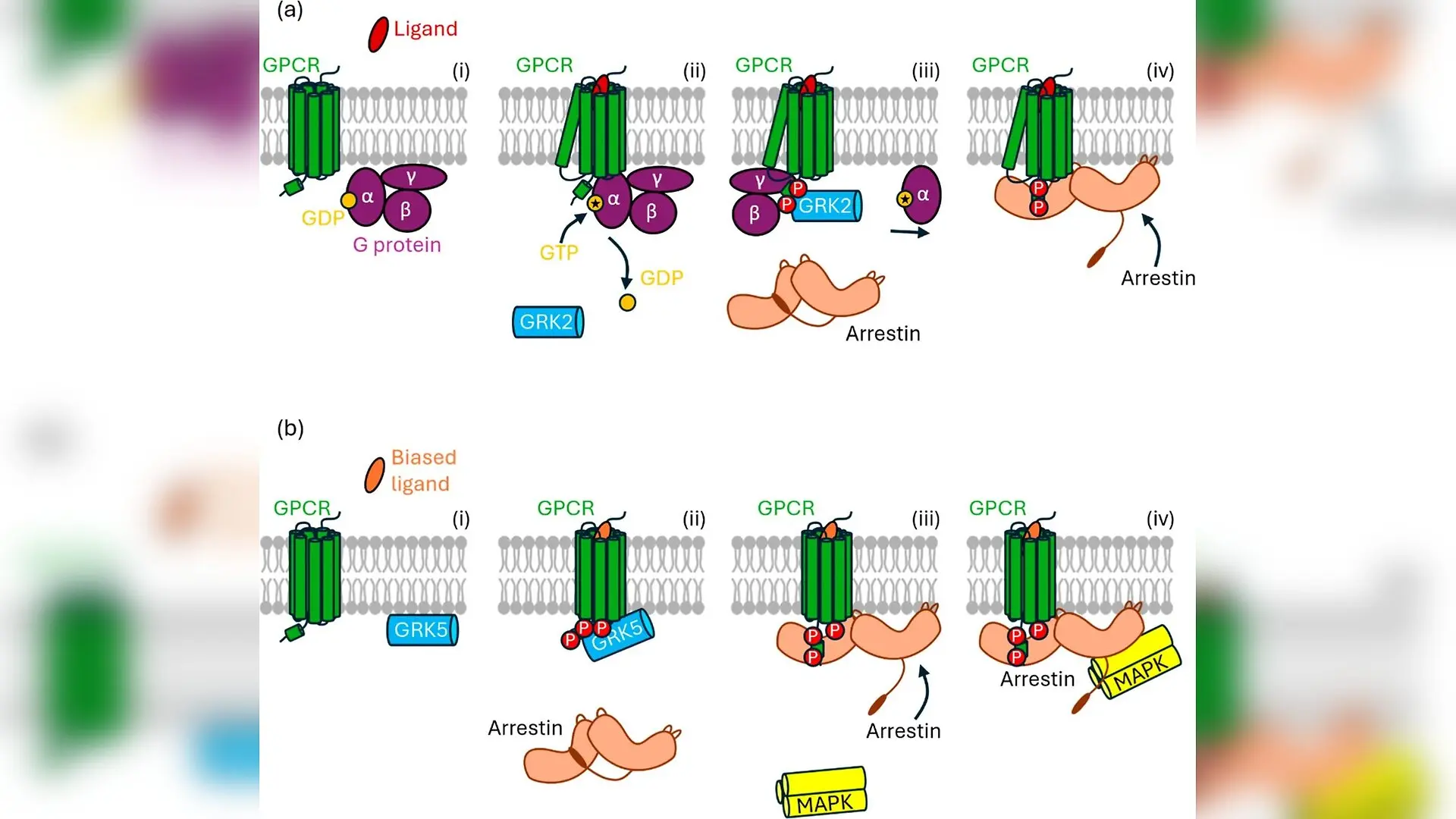
Investigating how cell surface receptors communicate with internal signaling molecules is fundamental to modern molecular biology, yet observing these events in real time within living cells remains extraordinarily difficult. G protein-coupled receptors represent one of the largest and most therapeutically vital classes of membrane proteins in human biology, mediating responses to everything from hormones to neurotransmitters. Despite decades of intense study, capturing the precise choreography of these proteins as they recruit downstream partners like arrestins has been hindered by instrumental drift and the physical limitations of microscope stages.
When an agonist ligand binds to a receptor, it triggers a cascade of intracellular events, including the movement of cytoplasmic proteins toward the plasma membrane. Documenting this translocation typically requires imaging both the membrane interface and deeper cellular cross sections alternately over extended periods. Conventional microscope hardware suffers from mechanical backlash, thermal fluctuations, and positioning hysteresis, making it nearly impossible to return to the exact same focal plane repeatedly without introducing massive experimental noise. Consequently, researchers have historically been forced to average measurements across dozens of different cells, smoothing over individual cellular behaviors and obscuring vital physiological variations.
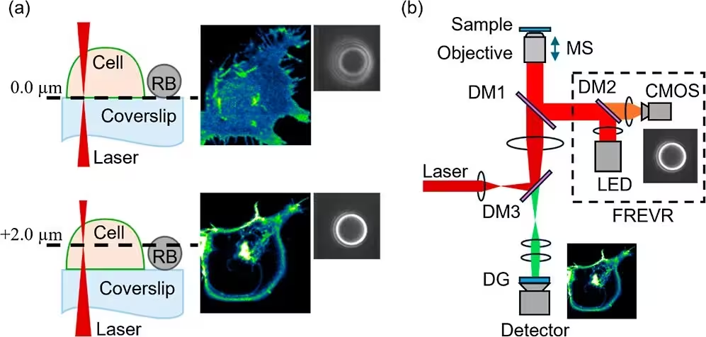
To overcome these barriers, a collaborative group of biophysicists and chemists published in The Journal of Physical Chemistry B detailing a novel optical configuration. Their approach integrates a specialized stabilization system known as Focal Readjustment for Enhanced Vertical Resolution, or FREVR, into a custom two-photon microspectroscope. Instead of relying on expensive specialized piezo stages, the team utilized amino-coated polystyrene reference beads attached to the coverslip as fiducial markers. A dedicated secondary optical pathway equipped with an LED and a CMOS camera continuously tracks these reference beads against a pre-recorded high-resolution lookup library, feeding real-time positional data directly to the microscope software.
Achieving Nanometer-Scale Focal Stability
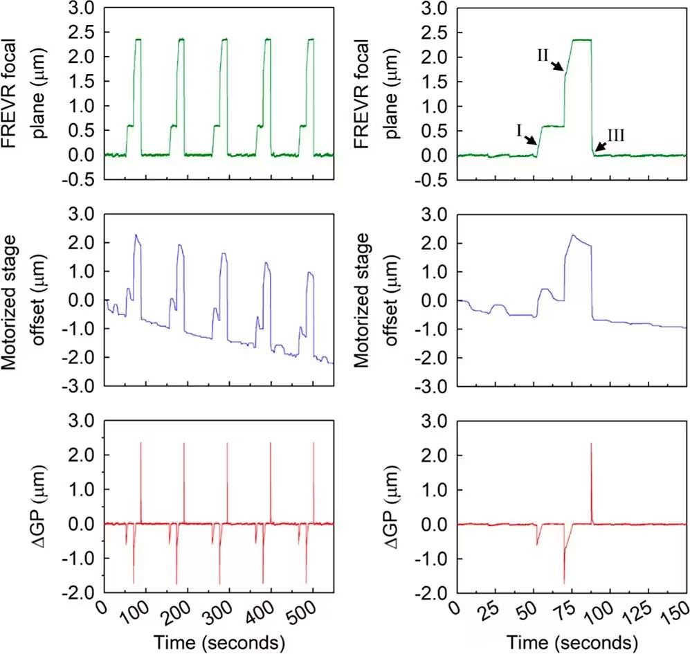
The implementation of this technology yielded exceptional stability during long-term imaging sessions. Benchmark tests demonstrated that the system maintains an axial positioning precision of better than twenty nanometers over extended experimental windows. When the motorized stage undershot commanded positions due to mechanical imperfections, the stabilization algorithm actively compensated in real time, neutralizing cumulative focal drift that would otherwise ruin multi-plane time-lapse datasets.
With this robust stabilization framework established, the researchers applied the setup to human embryonic kidney cells expressing fluorescently tagged muscarinic acetylcholine M2 receptors and arrestin-2. The experimental protocol involved alternating between two specific focal planes, the basolateral plasma membrane and a cross-sectional plane positioned over two micrometers deeper into the cellular interior. This bisectional strategy allowed the team to monitor receptor clustering and cytoplasmic protein recruitment simultaneously within the exact same cell.
Unveiling Receptor and Arrestin Dynamics
Following stimulation with the agonist carbachol, the imaging data revealed striking spatial and temporal reorganizations. At the basolateral membrane, receptors that were initially distributed uniformly began to coalesce into dense punctate structures resembling clathrin-coated pits. Simultaneously, cross-sectional views captured the progressive depletion of fluorescently tagged arrestin-2 from the cytoplasm as it migrated toward the inner leaflet of the plasma membrane.
Quantitative Single-Cell Insights
By coupling these imaging planes with advanced spectral unmixing algorithms and mathematical modeling, the investigators mapped protein concentrations quantitatively over time. The results showed that arrestin translocation and its colocalization with receptors at membrane puncta followed sigmoidal kinetics, occurring on tightly matched timescales. Because the stabilization system eliminated the need for population averaging, individual cell trajectories emerged with striking clarity, highlighting natural variations in cellular responsiveness.
The ability to access multiple focal planes with nanometer-scale precision opens new avenues for studying complex signaling networks. While the optical detection optics remain bound by traditional diffraction limits, the dramatic reduction in mechanical instability empowers researchers to investigate the stoichiometry, kinetics, and spatial organization of membrane-cytoplasm interactions with confidence.
The research was published in The journal of physical chemistry. B on October 8, 2026.
This article has been fact checked for accuracy, with information verified against reputable sources. Learn more about us and our editorial process.
Last reviewed on .
Article history
- Latest version
Reference(s)
- Killeen, Thomas D.., et al. “Precise Alternation between Image-Forming Sample Planes Enables Quantitative Monitoring of Receptor-Arrestin Interaction Dynamics at the Plasma Membrane of Live Cells.” The Journal of Physical Chemistry B, vol. 130, no. 40, September 24, 2026, pp. 10371-10382. American Chemical Society (ACS), doi: 10.1021/acs.jpcb.6c02313. <https://doi.org/10.1021/acs.jpcb.6c02313>.
Cite this page:
- Posted by John Williams