New Topological Framework Reveals How Atrial Fibrillation Waves Form and Terminate
By reducing complex cardiac arrhythmias into two fundamental wave archetypes and five basic events, scientists achieve a 100% success rate in virtual ablation simulations.
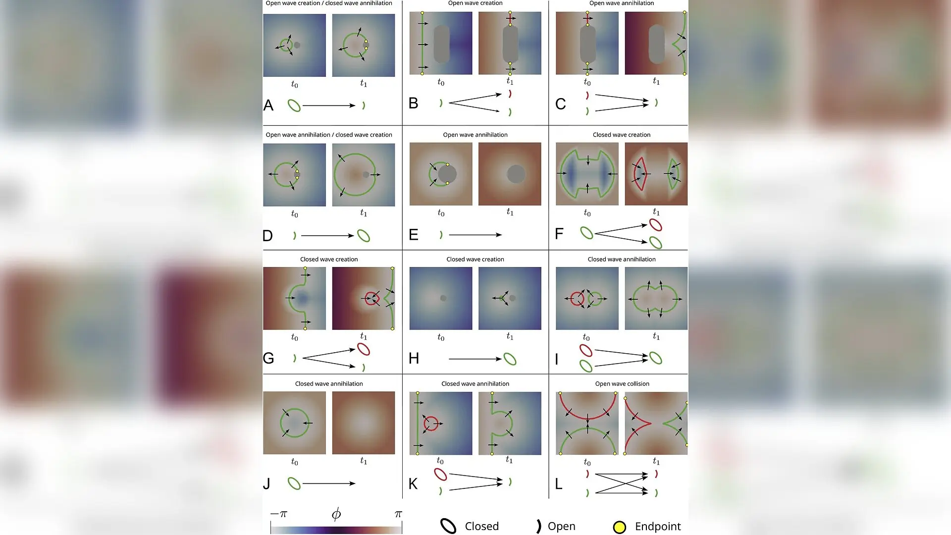
Atrial arrhythmias, most notably atrial fibrillation, present some of the most intricate spatiotemporal patterns in cardiac electrophysiology. For decades, researchers have debated the exact mechanisms that sustain persistent forms of the condition, putting forward competing theories such as the multiple wavelet hypothesis, the mother rotor hypothesis, and various anatomical or intramural reentry models. While these concepts have guided modern cardiology, fully characterizing the chaotic propagation of electrical signals across the heart tissue has remained an elusive goal. A new study published in Frontiers in Network Physiology now demonstrates that much of this apparent complexity stems from simple, underlying topological principles governing how electrical waves interact and evolve.
Simplifying Chaos Into Two Wave Archetypes
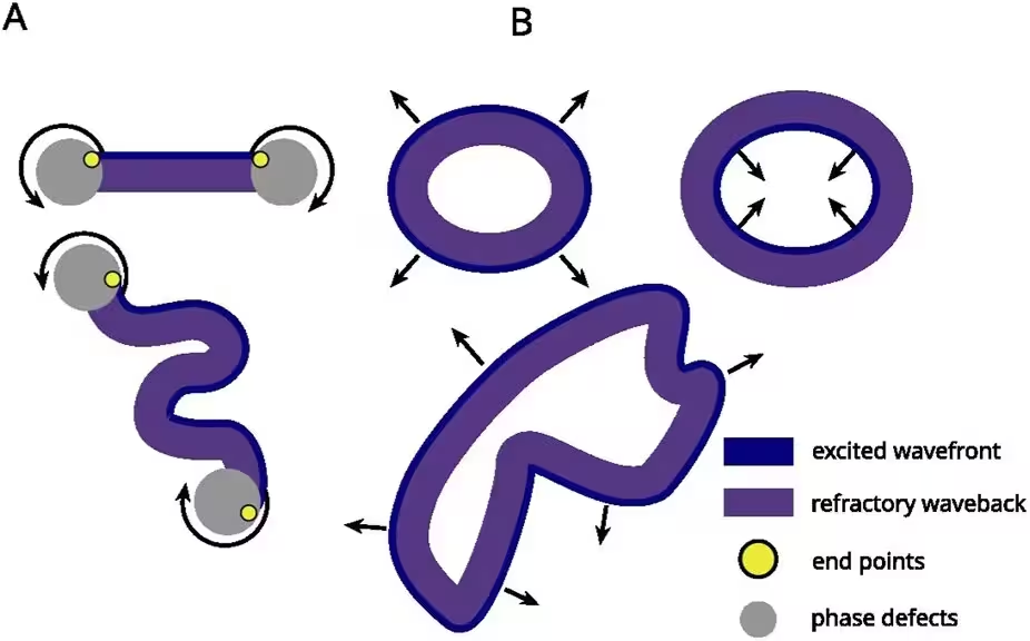
To cut through the complexity of fibrillatory conduction, the research team shifted their perspective from traditional tracking of localized rotation centers to a comprehensive analysis of wavefront topology. Working with single-atrium meshes modeled as two-dimensional surfaces with natural openings like valves and veins, the investigators established that all cardiac wavefronts belong to one of two fundamental categories: open waves and closed waves.
Open waves are line-like structures bounded by two endpoints that connect to anatomical or functional phase defects, such as scar tissue or refractory regions. These open structures tend to self-perpetuate and encompass phenomena such as rotors and meandering wavelets. In contrast, closed waves form continuous ring-like structures enclosing a region entirely, possessing no endpoints. While expanding closed waves eventually transition into shrinking waves and collapse, open waves require specific interactions to change their state.
Five Elementary Interaction Processes
Through detailed analysis of 600 patient-based computer simulations generated using the openCARP software platform, the authors mapped out how these wavefronts behave when they collide with each other or with anatomical obstacles. They discovered that all complex interactions reduce to five elementary processes:
- Open wave creation, where an obstacle splits a wave or converts a closed wave into an open structure.
- Open wave annihilation, where opposite endpoints collide and disappear, sometimes turning an open wave into a closed loop.
- Closed wave creation, arising from self-collisions or focal activity.
- Closed wave annihilation, occurring when closed loops shrink, merge, or interact with open waves.
- Open wave collision, where two open waves meet frontally and generate two new open waves without altering the net wave count.
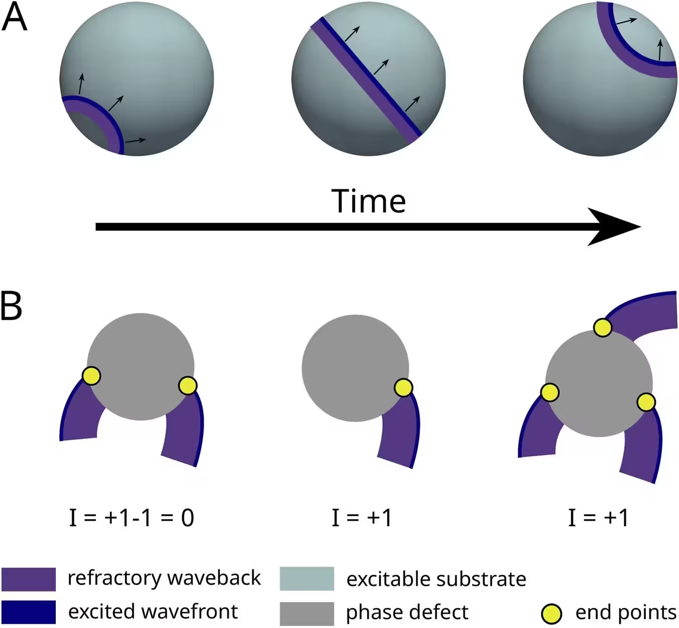
Every single event observed across the extensive simulation dataset conformed strictly to these topological rules. Furthermore, by calculating rotational indices around phase defects rather than traditional phase singularities, the team confirmed that the total topological charge across the compact atrial surface consistently summed to zero, perfectly satisfying the index theorem.
Virtual Ablation and Future Clinical Implications
The practical payoff of this topological classification emerged when the researchers tested a novel virtual ablation strategy. Instead of conventional ablation lines designed merely to block local pathways, the new approach identified all phase defects and targeted the endpoints of open waves, effectively connecting them to common boundaries to force rapid self-annihilation.
When evaluated across the simulation cohort, this topology-informed intervention achieved a 100 percent success rate in eliminating rotational activity and establishing electrical silence before the end of the simulation runs. Although implementing instantaneous, dynamic ablation lines in real-world clinical settings presents significant technological hurdles, the framework provides a powerful new lens for understanding arrhythmia maintenance. By identifying patient-specific tissue substrates prone to generating non-recombining open waves, cardiologists may one day be able to design more precise, individualized ablation strategies that permanently silence persistent atrial fibrillation.
The research was published in Frontiers in network physiology on January 1, 2026.
This article has been fact checked for accuracy, with information verified against reputable sources. Learn more about us and our editorial process.
Last reviewed on .
Article history
- Latest version
Reference(s)
- Bezerra, Arthur S.., et al. “Open wavefront ablation terminates rotor-driven atrial fibrillation.” Frontiers in Network Physiology, vol. 6, October 8, 2026 Frontiers Media SA, doi: 10.3389/fnetp.2026.1940428. <https://doi.org/10.3389/fnetp.2026.1940428>.
Cite this page:
- Posted by John Williams